QUESTION IMAGE
Question
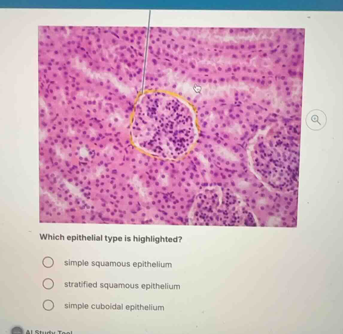
which epithelial type is highlighted?
simple squamous epithelium
stratified squamous epithelium
simple cuboidal epithelium
Identify the highlighted structure
The image shows a histological section of the kidney cortex. The pointer and the yellow highlighted circle outline the parietal layer of the glomerular capsule (Bowman's capsule), which surrounds the glomerulus.
Analyze the tissue type
The parietal layer of the glomerular capsule is composed of a single layer of flat, scale-like cells. This specific tissue structure is classified as Simple Squamous Epithelium Locations.
Compare with other options
- Simple Squamous Epithelium: This is the correct tissue type forming the thin outer wall (parietal layer) of the glomerular capsule.
- Stratified Squamous Epithelium: This consists of multiple layers of flat cells, typically found in protective barriers like the skin or esophagus, not here.
- Simple Cuboidal Epithelium: While present in the surrounding renal tubules (visible as the round structures with round nuclei surrounding the glomerulus), it is not the tissue highlighted by the yellow outline of the capsule wall.

Snap & solve any problem in the app
Get step-by-step solutions on Sovi AI
Photo-based solutions with guided steps
Explore more problems and detailed explanations
- simple squamous epithelium (Correct answer)
- stratified squamous epithelium
- simple cuboidal epithelium