QUESTION IMAGE
Question
Question was provided via image upload.
🆕 New Concept Discovered: Histological Identification of Connective Tissues
Recognizing tissue types by fiber arrangement and cell density.
Step 1: Analyze the visual characteristics of the tissue
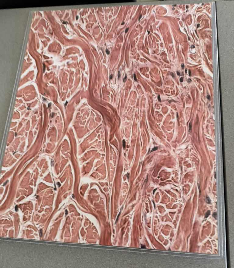
Looking closely at the micrograph, we can observe several key histological features:
- Fibers: There are thick, wavy, pink-staining bundles running in many different directions. These are collagen fibers.
- Arrangement: The collagen bundles do not run parallel to one another; instead, they form a random, interwoven, three-dimensional network.
- Cells: There are scattered, dark-stained nuclei belonging to fibroblasts, which are the cells that produce these fibers.
- Ground Substance: There is very little open space (ground substance) compared to the dense volume of collagen fibers.
Step 2: Correlate features with tissue types
Let's compare these observations to the main categories of connective tissue:
- Loose (Areolar) Connective Tissue: Contains a lot of open space (ground substance) with thin, loosely scattered fibers. This tissue is far too packed with thick fibers to be loose connective tissue.
- Dense Regular Connective Tissue: Contains thick collagen fibers, but they are arranged in parallel, orderly bundles (like in tendons or ligaments). The fibers here are highly disorganized and run in multiple directions.
- Dense Irregular Connective Tissue: Characterized by a dense packing of thick collagen fiber bundles that are arranged randomly and irregularly, allowing the tissue to resist tension applied from many different directions. This perfectly matches the image.

Snap & solve any problem in the app
Get step-by-step solutions on Sovi AI
Photo-based solutions with guided steps
Explore more problems and detailed explanations
Dense irregular connective tissue