QUESTION IMAGE
Question
Question was provided via image upload.
🆕 New Concept Discovered: Histological Identification of Tissues and Cells
Recognizing tissue types and blood cells under a microscope.
Step 1: Analyze the bottom image
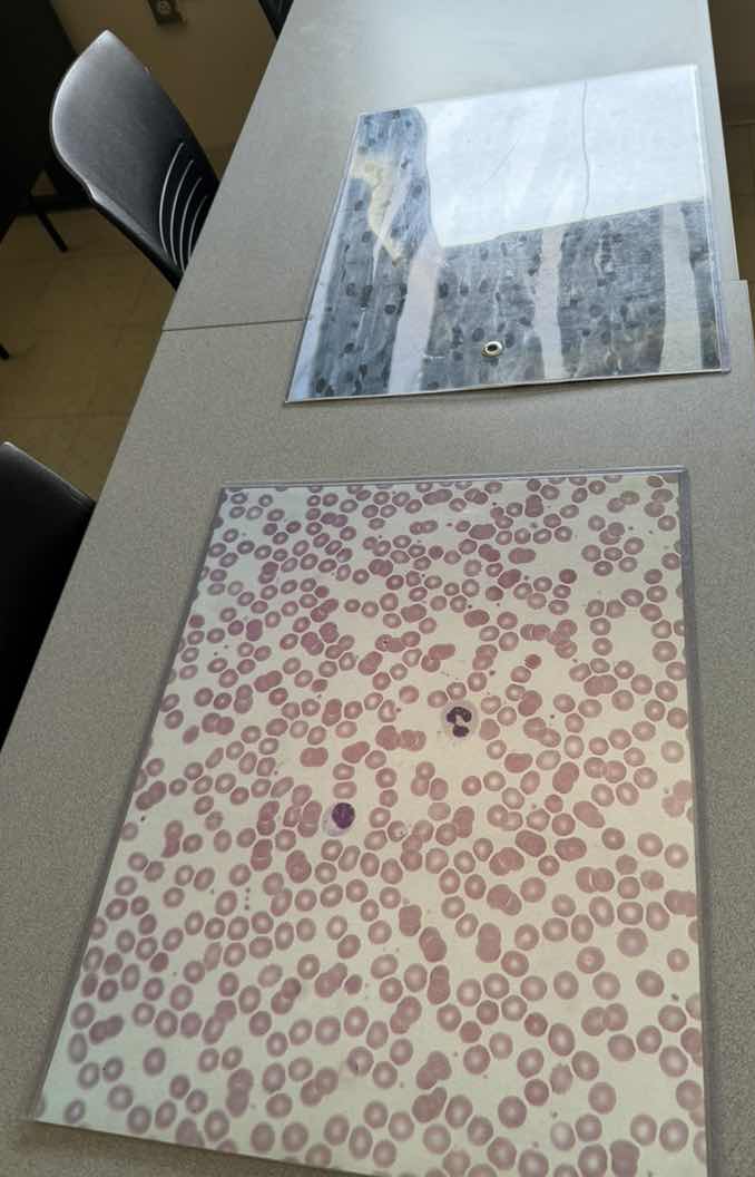
The bottom image shows a microscopic view of a blood smear:
- The vast majority of the cells are small, pinkish-red, circular, and lack a nucleus (biconcave discs). These are erythrocytes (red blood cells).
- There are a couple of larger cells with distinct, dark purple-stained nuclei. These are leukocytes (white blood cells). Specifically, the one near the center has a multi-lobed nucleus characteristic of a neutrophil, and the one slightly lower and to the left has a large, dense nucleus characteristic of a lymphocyte.
Step 2: Analyze the top image
The top image shows a microscopic view of a tissue section:
- It displays long, parallel, cylindrical fibers running vertically.
- There are distinct dark nuclei located at the periphery of these fibers.
- Cross-striations (alternating light and dark bands) are visible running perpendicular to the length of the fibers.
- These features are highly characteristic of skeletal muscle tissue (striated, multinucleated fibers with peripheral nuclei).

Snap & solve any problem in the app
Get step-by-step solutions on Sovi AI
Photo-based solutions with guided steps
Explore more problems and detailed explanations
- Bottom Image: Blood smear showing erythrocytes (red blood cells) and leukocytes (white blood cells, including a neutrophil and a lymphocyte).
- Top Image: Skeletal muscle tissue (showing striated muscle fibers with peripheral nuclei).