QUESTION IMAGE
Question
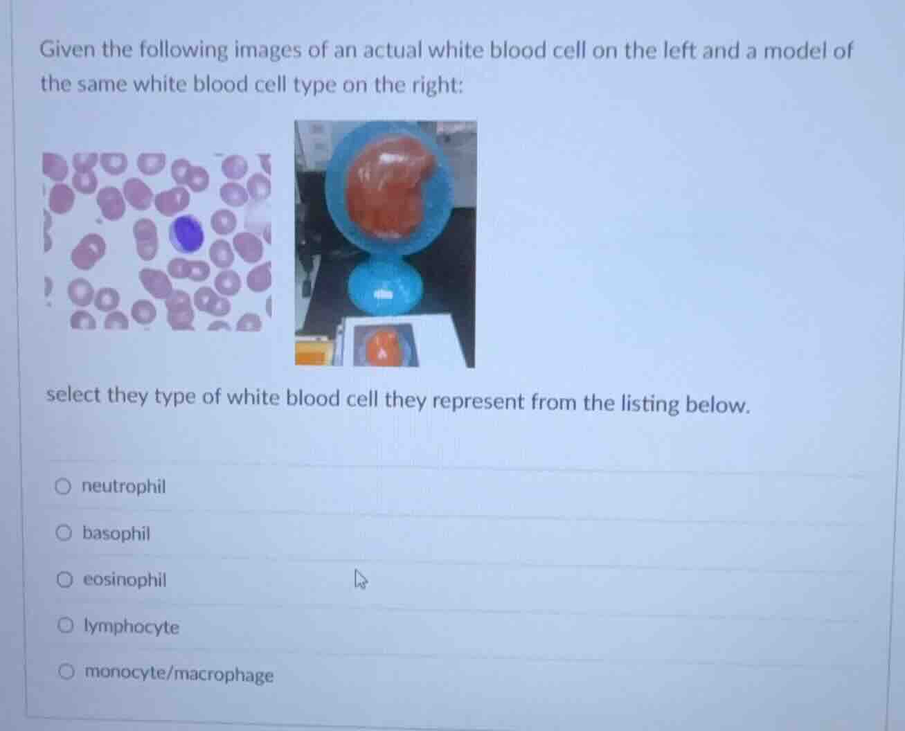
given the following images of an actual white blood cell on the left and a model of the same white blood cell type on the right:
select they type of white blood cell they represent from the listing below.
neutrophil
basophil
eosinophil
lymphocyte
monocyte/macrophage
Analyze the microscopic image
The image on the left shows a blood smear. Among the red blood cells, there is a large white blood cell with a large, indented, kidney-shaped (or horseshoe-shaped) nucleus. The cytoplasm is abundant and lacks prominent, brightly colored granules, which is characteristic of an agranulocyte.
Analyze the physical model
The model on the right represents the same cell type. It features a large, distinct kidney-shaped nucleus (colored reddish-brown) occupying a significant portion of the cell, which is a classic diagnostic feature used in anatomy and physiology labs to represent a monocyte.
Compare with leukocyte types
- Neutrophils have multi-lobed nuclei (3 to 5 lobes) and pale granules.
- Basophils have dark blue/purple granules that often obscure the nucleus.
- Eosinophils have bilobed nuclei and bright red/orange granules.
- Lymphocytes are smaller, with a round, dense nucleus that fills most of the cell.
- Monocytes are the largest leukocytes, featuring a characteristic kidney- or U-shaped nucleus and abundant agranular cytoplasm.
Match with options
The morphological features of both the real cell and the model perfectly match the characteristics of a monocyte/macrophage.

Snap & solve any problem in the app
Get step-by-step solutions on Sovi AI
Photo-based solutions with guided steps
Explore more problems and detailed explanations
- neutrophil
- basophil
- eosinophil
- lymphocyte
- monocyte/macrophage (Correct answer)