QUESTION IMAGE
Question
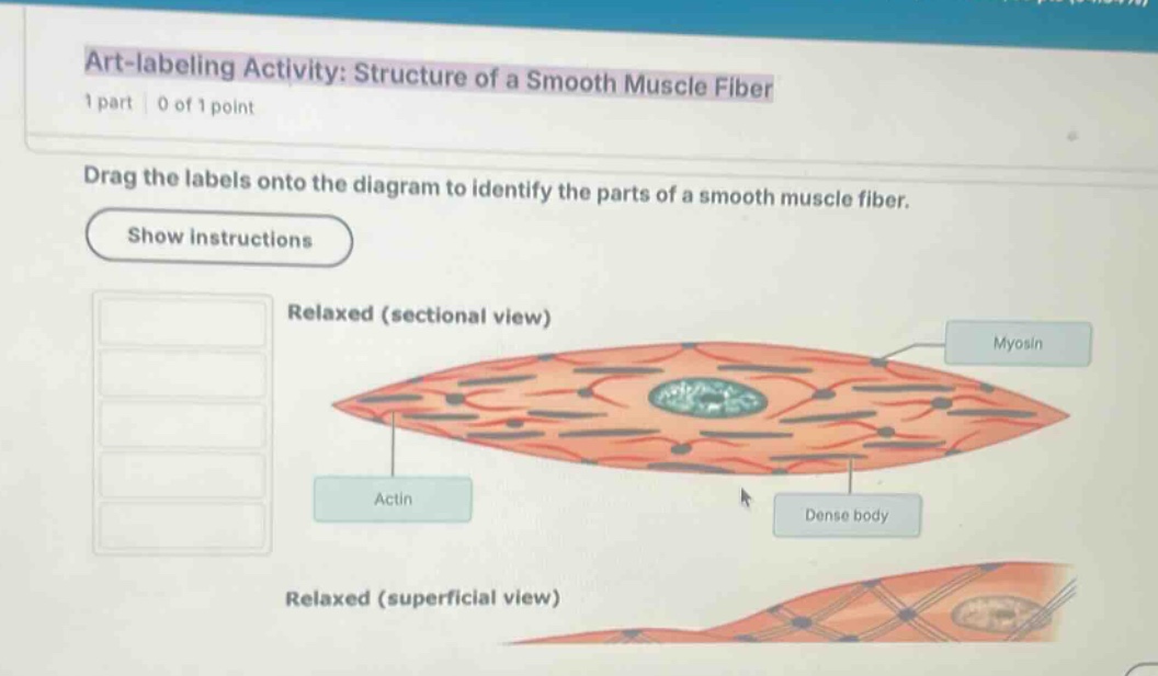
art-labeling activity: structure of a smooth muscle fiber
1 part | 0 of 1 point
drag the labels onto the diagram to identify the parts of a smooth muscle fiber.
show instructions
relaxed (sectional view)
actin
myosin
dense body
relaxed (superficial view)
🆕 New Concept Discovered: Smooth Muscle Microanatomy
Understanding how non-striated muscle cells organize contractile proteins.
Step 1: Identify the visible labels
The image shows a diagram of a smooth muscle fiber in a relaxed state (sectional view and superficial view).
Three labels are already placed on the sectional view:
- Actin: Points to the thin filaments (red lines).
- Myosin: Points to the thick filaments (darker, thicker bars).
- Dense body: Points to the dark circular structures anchoring the thin filaments (analogous to Z-discs in striated muscle).
Step 2: Determine the missing labels
In standard anatomy art-labeling activities for the "Structure of a Smooth Muscle Fiber" (typically from Pearson/Marieb Human Anatomy & Physiology), the labels to be dragged onto the diagram generally include:
- Sarcolemma (the plasma membrane of the muscle cell)
- Nucleus (the central, single oval structure in the middle of the cell)
- Intermediate filaments (the non-contractile cytoskeletal elements connecting the dense bodies)
- Caveolae (pouch-like infoldings of the sarcolemma that sequester calcium)
- Dense plaques (or attachment plaques, anchoring dense bodies to the sarcolemma)
Looking at the sectional view:
- The large, central, textured oval structure in the middle of the cell is the Nucleus.
- The outer boundary membrane of the cell is the Sarcolemma.
- The network of structural lines running between dense bodies represents Intermediate filaments.

Snap & solve any problem in the app
Get step-by-step solutions on Sovi AI
Photo-based solutions with guided steps
Explore more problems and detailed explanations
Based on standard anatomical diagrams of a smooth muscle fiber, the remaining labels to place on the diagram are:
- Nucleus (points to the central blue/green textured organelle)
- Sarcolemma (points to the outer cell membrane)
- Intermediate filaments (points to the cytoskeletal lattice lines connecting the dense bodies)
- Caveolae (points to the tiny indentations along the sarcolemma membrane)