QUESTION IMAGE
Question
apr - cardiovascular system
activity 10: quiz
module: cardiovascular
topic: structures / type: multiple choice (mc)
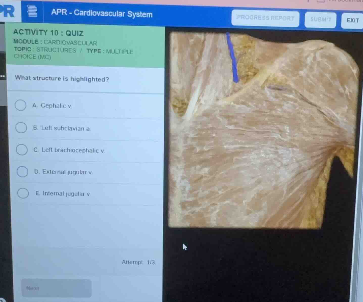
what structure is highlighted?
a. cephalic v.
b. left subclavian a.
c. left brachiocephalic v.
d. external jugular v.
e. internal jugular v.
attempt 1/3
Identify the anatomical region and orientation
The image shows a cadaveric dissection of the anterior chest and neck region. The clavicle is visible, with the pectoralis major muscle inferior to it. Superior to the clavicle, we can observe the structures of the lower neck.
Analyze the highlighted structure
Using the Venous Anatomy knowledge point
The highlighted blue structure is a superficial vein running vertically down the lateral aspect of the neck, crossing superficial to the sternocleidomastoid muscle. It descends toward the subclavian vein.
Differentiate between the options
- Cephalic v.: Runs in the deltopectoral groove between the deltoid and pectoralis major muscles, which is lateral and inferior to this region.
- Left subclavian a.: A deep artery located posterior to the clavicle, not a superficial vertical neck structure.
- Left brachiocephalic v.: Located deep within the superior mediastinum, formed by the union of the internal jugular and subclavian veins.
- Internal jugular v.: A deep vein running adjacent to the common carotid artery inside the carotid sheath, deep to the sternocleidomastoid muscle.
- External jugular v.: A superficial vein of the neck that runs vertically down, superficial to the sternocleidomastoid muscle, to drain into the subclavian vein. This matches the highlighted structure perfectly.

Snap & solve any problem in the app
Get step-by-step solutions on Sovi AI
Photo-based solutions with guided steps
Explore more problems and detailed explanations
- (A) Cephalic v.
- (B) Left subclavian a.
- (C) Left brachiocephalic v.
- (D) External jugular v. (Correct answer)
- (E) Internal jugular v.