QUESTION IMAGE
Question
module: nervous
topic: structures / type: combination
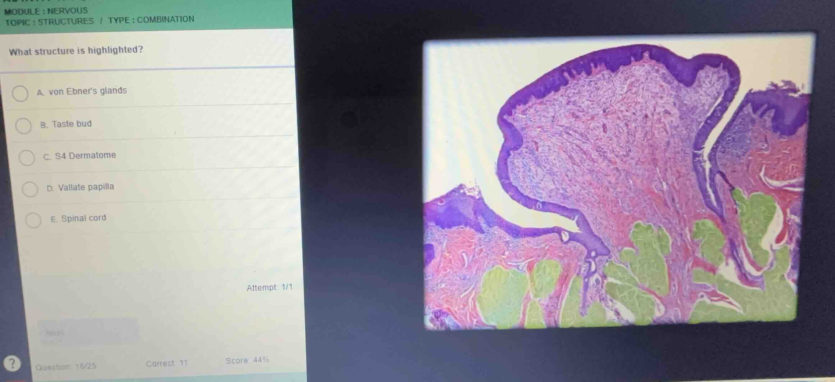
what structure is highlighted?
a. von ebners glands
b. taste bud
c. s4 dermatome
d. vallate papilla
e. spinal cord
Identify the histological specimen
Using the Tongue Histology and Vallate Papilla knowledge points
The image shows a high-power histological section of a tongue papilla, specifically a vallate papilla (circumvallate papilla), characterized by its large dome-shaped structure surrounded by a deep circular furrow (trench).
Analyze the highlighted region
Using the Tongue Histology and Taste Buds knowledge points
The green-shaded structures located deep in the lamina propria beneath the trench of the vallate papilla are serous salivary glands. These are von Ebner's glands (gustatory glands), which secrete a watery fluid into the cleft to flush out food particles and allow taste buds to register new gustatory stimuli.
Correlate with options
Using the Tongue Histology knowledge point
- A. von Ebner's glands: Correct, as these are the serous glands highlighted in green at the base of the furrow.
- B. Taste bud: Incorrect; taste buds are located in the lateral epithelial walls of the papilla, not deep in the connective tissue.
- C. S4 Dermatome: Incorrect; unrelated anatomical structure.
- D. Vallate papilla: Incorrect; the papilla itself is the entire dome-shaped mucosal projection, not just the green-highlighted basal glands.
- E. Spinal cord: Incorrect; unrelated tissue.

Snap & solve any problem in the app
Get step-by-step solutions on Sovi AI
Photo-based solutions with guided steps
Explore more problems and detailed explanations
- (A) von Ebner's glands (Correct answer)
- (B) Taste bud
- (C) S4 Dermatome
- (D) Vallate papilla
- (E) Spinal cord