QUESTION IMAGE
Question
Question was provided via image upload.
Identify the anatomical structure
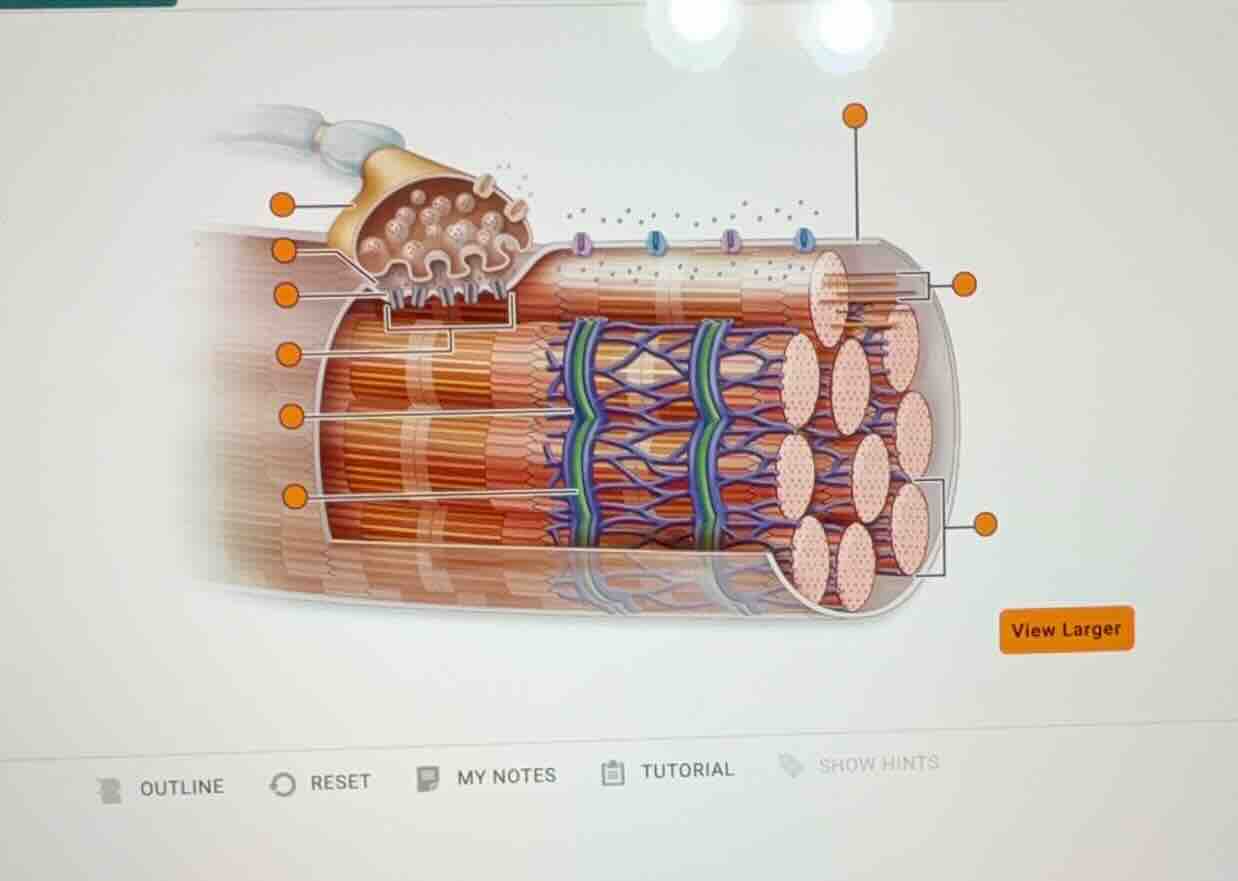
This diagram illustrates a skeletal muscle fiber (cell) and its innervation by a motor neuron at the neuromuscular junction.
Analyze the labeled components
Let's trace the leader lines with orange circular terminals from top to bottom on the left, and then on the right:
Left side labels (from top to bottom):
- Synaptic terminal (Axon terminal): The bulbous distal end of the motor neuron containing synaptic vesicles.
- Synaptic cleft: The narrow space between the synaptic terminal and the muscle fiber sarcolemma.
- Junctional folds (Motor end plate): The folded region of the sarcolemma directly under the synaptic terminal.
- Myofibril: The cylindrical contractile organelles packed inside the muscle fiber.
- Sarcoplasmic reticulum (SR): The blue/purple web-like network surrounding each myofibril that stores calcium ions.
- Transverse tubule (T-tubule): The green-colored invaginations of the sarcolemma that run deep into the fiber.
Right side labels (from top to bottom):
- Sarcolemma: The plasma membrane of the muscle fiber (indicated by the top-right leader line pointing to the outer membrane).
- Mitochondria: The elongated organelles situated between myofibrils (indicated by the middle-right leader line pointing to the orange oval structures).
- Myofibrils (cross-section): The group of circular profiles representing the cut ends of the myofibrils (indicated by the bracketed bottom-right leader line).
Provide a comprehensive guide
Since the interactive labels are blank, we will provide the correct anatomical term for each pointer so you can easily drag and drop or fill in the answers.

Snap & solve any problem in the app
Get step-by-step solutions on Sovi AI
Photo-based solutions with guided steps
Explore more problems and detailed explanations
Based on the anatomical diagram of a skeletal muscle fiber and the neuromuscular junction, here are the correct labels corresponding to each pointer:
Left Side (Top to Bottom)
- First pointer: Synaptic terminal (Axon terminal of motor neuron)
- Second pointer: Synaptic cleft
- Third pointer: Junctional folds (Motor end plate of sarcolemma)
- Fourth pointer: Myofibril
- Fifth pointer: Sarcoplasmic reticulum
- Sixth pointer: T-tubule (Transverse tubule)
Right Side (Top to Bottom)
- Top pointer: Sarcolemma
- Middle pointer: Mitochondrion
- Bottom bracketed pointer: Myofibrils (cross-section)