QUESTION IMAGE
Question
Question was provided via image upload.
Identify the anatomical structures
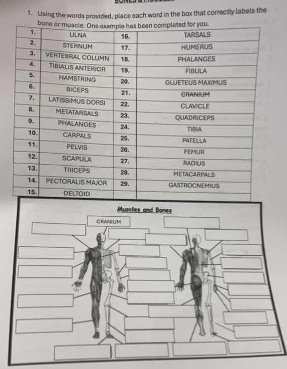
The task is to label the muscles and bones of the human body using the 29 provided terms. The diagram shows an anterior view (left figure) and a posterior view (right figure), with one side showing muscles and the other side showing bones. Let's match each term to its correct anatomical location.
Analyze the anterior view (left figure)
- Cranium: Already labeled at the top of the skull.
- Deltoid: Shoulder muscle.
- Pectoralis Major: Chest muscle.
- Biceps: Front upper arm muscle.
- Radius & Ulna: Forearm bones. The radius is lateral (thumb side), and the ulna is medial (pinky side).
- Carpals, Metacarpals, Phalanges: Hand bones. Carpals are wrist, metacarpals are palm, phalanges are fingers.
- Sternum: Breastbone in the center of the chest.
- Ribs / Vertebral Column: Central axial skeleton.
- Pelvis: Hip bone structure.
- Femur: Thigh bone.
- Patella: Kneecap.
- Tibia & Fibula: Shin bones. Tibia is the larger medial bone; fibula is the thinner lateral bone.
- Tarsals, Metatarsals, Phalanges: Foot bones. Tarsals are ankle, metatarsals are sole, phalanges are toes.
- Quadriceps: Front thigh muscle.
- Tibialis Anterior: Front shin muscle.
Analyze the posterior view (right figure)
- Trapezius / Latissimus Dorsi: Back muscles. Latissimus dorsi is the large lower back muscle.
- Triceps: Back upper arm muscle.
- Gluteus Maximus: Buttock muscle.
- Hamstring: Back thigh muscle.
- Gastrocnemius: Calf muscle.
- Vertebral Column: Spine visible from the back.
- Scapula: Shoulder blade.
- Clavicle: Collarbone (visible near the top shoulder area).
- Humerus: Upper arm bone.
Match labels to diagram pointers
Let's systematically list the correct anatomical terms corresponding to the standard locations indicated by the pointer lines in such worksheets:
- Head/Skull: Cranium (Bone - labeled)
- Shoulder Muscle: Deltoid
- Chest Muscle: Pectoralis Major
- Upper Arm Muscle (Front): Biceps
- Upper Arm Muscle (Back): Triceps
- Upper Arm Bone: Humerus
- Forearm Bone (Lateral): Radius
- Forearm Bone (Medial): Ulna
- Wrist Bones: Carpals
- Hand Bones (Palm): Metacarpals
- Finger/Toe Bones: Phalanges
- Breastbone: Sternum
- Shoulder Blade: Scapula
- Collarbone: Clavicle
- Spine: Vertebral Column
- Hip Bone: Pelvis
- Thigh Bone: Femur
- Kneecap: Patella
- Shin Bone (Inner/Larger): Tibia
- Shin Bone (Outer/Smaller): Fibula
- Ankle Bones: Tarsals
- Foot Bones (Sole): Metatarsals
- Thigh Muscle (Front): Quadriceps
- Thigh Muscle (Back): Hamstring
- Buttock Muscle: Gluteus Maximus
- Back Muscle: Latissimus Dorsi
- Calf Muscle: Gastrocnemius
- Shin Muscle (Front): Tibialis Anterior

Snap & solve any problem in the app
Get step-by-step solutions on Sovi AI
Photo-based solutions with guided steps
Explore more problems and detailed explanations
Based on the provided word bank and standard anatomical diagrams of the human muscular and skeletal systems, here is the identification of each term to help you fill in the blanks on your worksheet:
Bones (Skeletal System)
- Cranium: Skull (already completed as the example)
- Clavicle: Collarbone, located at the top of the chest/shoulder
- Scapula: Shoulder blade, located on the upper back
- Sternum: Breastbone, located in the center of the chest
- Humerus: Upper arm bone
- Radius: Forearm bone on the thumb side (lateral)
- Ulna: Forearm bone on the pinky side (medial)
- Carpals: Wrist bones
- Metacarpals: Bones of the palm of the hand
- Phalanges: Bones of the fingers and toes
- Vertebral Column: Spine/backbone running down the center of the back
- Pelvis: Hip bone structure
- Femur: Thigh bone (the longest bone in the body)
- Patella: Kneecap
- Tibia: Shinbone (the larger, inner bone of the lower leg)
- Fibula: Calf bone (the smaller, outer bone of the lower leg)
- Tarsals: Ankle bones
- Metatarsals: Bones of the sole of the foot
Muscles (Muscular System)
- Deltoid: Shoulder muscle
- Pectoralis Major: Large chest muscle
- Biceps: Muscle on the front of the upper arm
- Triceps: Muscle on the back of the upper arm
- Latissimus Dorsi: Large, flat muscle covering the lower back
- Gluteus Maximus: Large buttock muscle
- Quadriceps: Large muscle group on the front of the thigh
- Hamstring: Muscle group on the back of the thigh
- Gastrocnemius: Calf muscle on the back of the lower leg
- Tibialis Anterior: Muscle on the front of the shin