QUESTION IMAGE
Question
Question was provided via image upload.
Identify the first histological image
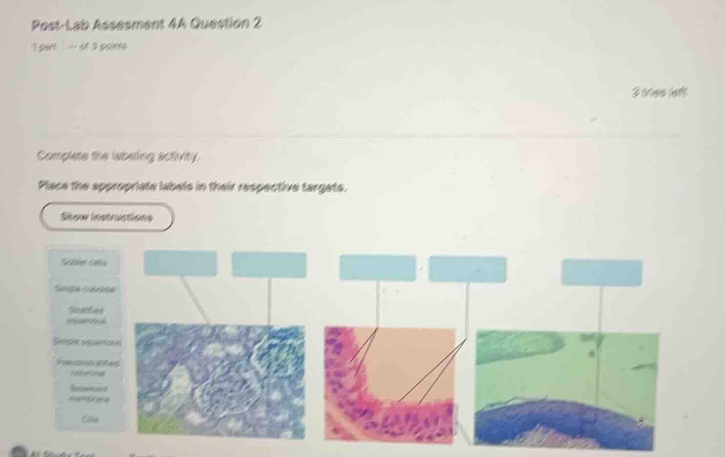
The first image on the left shows a section of the kidney cortex, specifically focusing on a renal corpuscle (glomerulus) surrounded by renal tubules.
- The left pointer points to the simple cuboidal epithelium lining the renal tubules.
- The right pointer points to the simple squamous epithelium forming the parietal layer of the glomerular capsule (Bowman's capsule).
- We apply our understanding of Simple Cuboidal Epithelium and Simple Squamous Epithelium Locations to identify these structures.
Identify the second histological image
The middle image shows a section of pseudostratified columnar epithelium, typically found in the respiratory tract (trachea).
- The left pointer points to the cilia on the apical surface of the cells.
- The right pointer points to the goblet cells interspersed among the columnar epithelial cells.
Identify the third histological image
The rightmost image shows a high-magnification view of an epithelial layer resting on underlying connective tissue.
- The pointer points to the thin, extracellular layer separating the epithelium from the connective tissue, which is the basement membrane.

Snap & solve any problem in the app
Get step-by-step solutions on Sovi AI
Photo-based solutions with guided steps
Explore more problems and detailed explanations
- First Image (Left, Kidney Tubules & Glomerulus):
- Left label: Simple cuboidal
- Right label: Simple squamous
- Second Image (Middle, Trachea/Respiratory Epithelium):
- Left label: Cilia
- Right label: Goblet cells
- Third Image (Right, Epithelial Boundary):
- Label: Basement membrane