QUESTION IMAGE
Question
Question was provided via image upload.
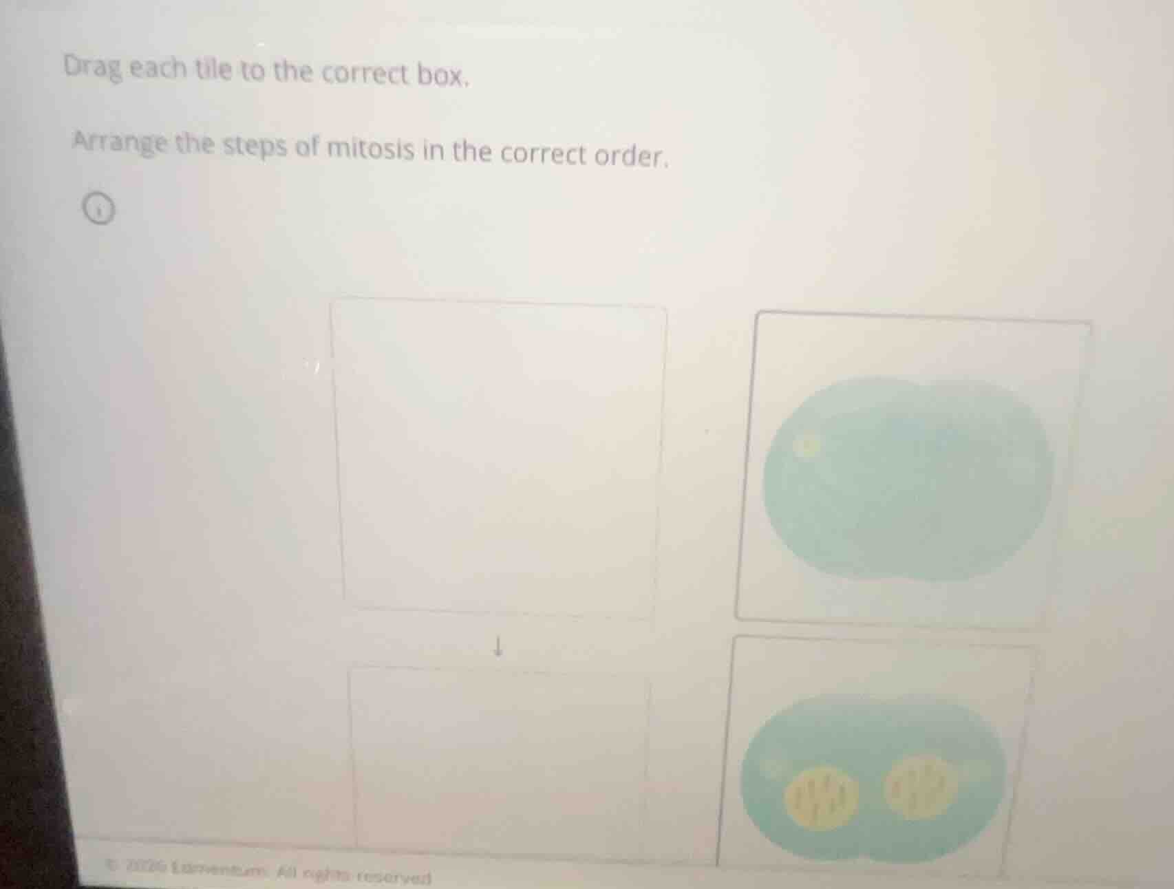
Identify the stages shown in the images
The image asks to arrange two steps of mitosis/cell division in the correct chronological order.
- The top-right image shows a cell undergoing cytokinesis where the cytoplasm is pinching in the middle (cleavage furrow), but the nuclear envelopes have not fully reformed or the genetic material is not clearly separated into distinct, organized nuclei yet.
- The bottom-right image shows a later stage of telophase/cytokinesis where two distinct, fully formed nuclei (yellow circles with chromatin/chromosomes inside) are visible within the pinching cell, preparing to complete division into two separate daughter cells.
Determine the chronological sequence
During cell division, the separation of genetic material and the initial constriction of the cell membrane occur first, followed by the complete reformation of the two distinct nuclei inside the dividing daughter cells before they fully separate.
- Step 1: The cell begins to pinch in the middle (top-right image).
- Step 2: Two distinct nuclei reform within the pinching cell structure (bottom-right image).

Snap & solve any problem in the app
Get step-by-step solutions on Sovi AI
Photo-based solutions with guided steps
Explore more problems and detailed explanations
To arrange the steps of mitosis in the correct order from top to bottom:
- First Box (Top): The image showing the cell pinching in the middle without fully reformed distinct nuclei (top-right tile).
- Second Box (Bottom): The image showing the pinching cell with two clearly visible, reformed yellow nuclei (bottom-right tile).