QUESTION IMAGE
Question
Question was provided via image upload.
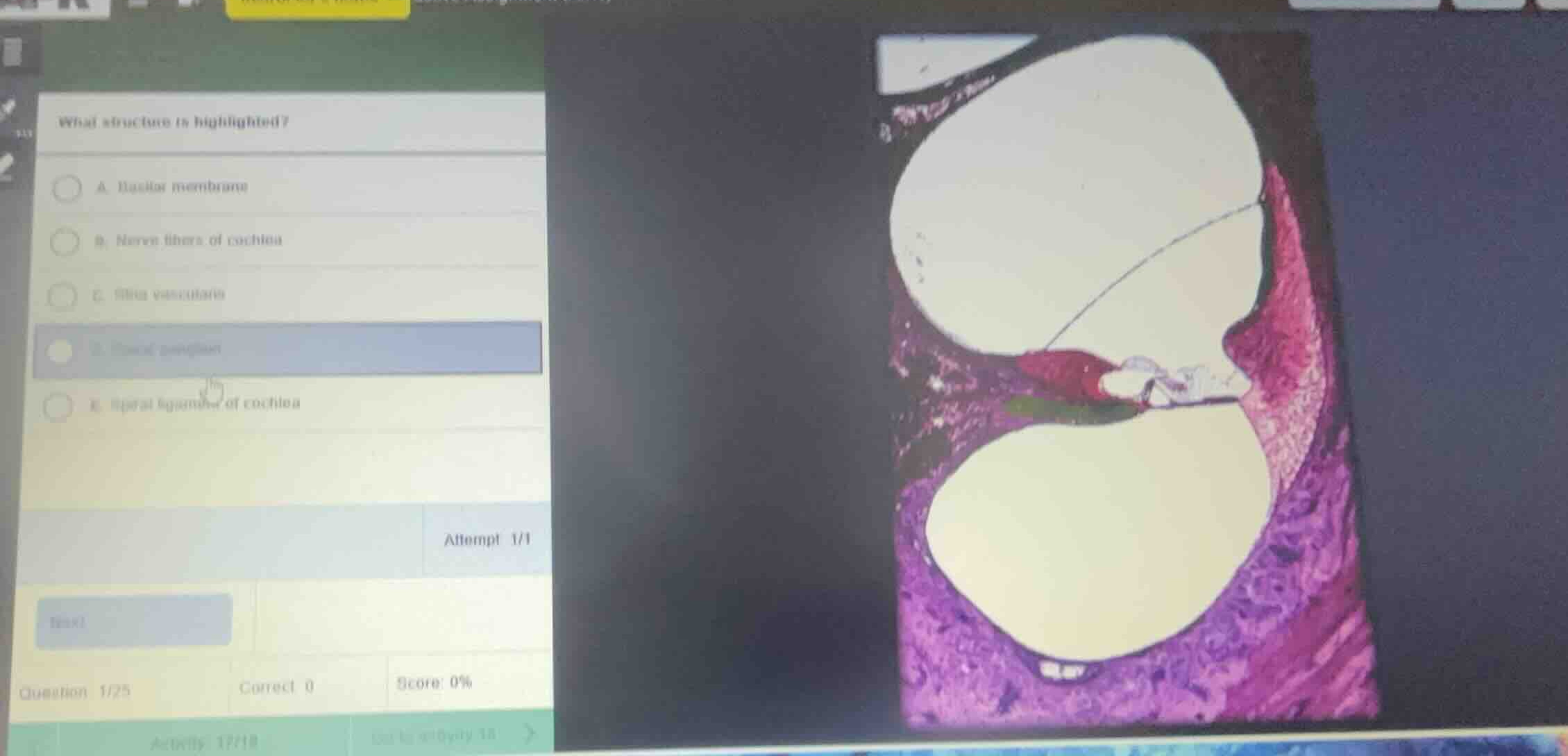
Identify the highlighted histological structure
Using the Cochlea Histology knowledge point
- The provided histological section shows a cross-section of a turn of the cochlea.
- The cochlear duct (scala media) is the middle triangular space, bounded superiorly by the vestibular membrane (Reissner's membrane) and inferiorly by the basilar membrane.
- The green highlighted region is located directly beneath the organ of Corti, forming the floor of the scala media.
- This specific floor structure supporting the sensory hair cells is the basilar membrane.
Match with the multiple-choice options
- Option A: Basilar membrane
- Option B: Nerve fibers of cochlea
- Option C: Stria vascularis
- Option D: Spiral ganglion
- Option E: Spiral ligament of cochlea
- The green highlighted structure corresponds to Option A, the basilar membrane.

Snap & solve any problem in the app
Get step-by-step solutions on Sovi AI
Photo-based solutions with guided steps
Explore more problems and detailed explanations
- (A) Basilar membrane (Correct answer)
- (B) Nerve fibers of cochlea
- (C) Stria vascularis
- (D) Spiral ganglion
- (E) Spiral ligament of cochlea