QUESTION IMAGE
Question
a&p flix activity: excitation-contraction coupling
called excitation-contraction coupling. this activity will test your understanding of the sequence of events that occur during excitation-contraction coupling.
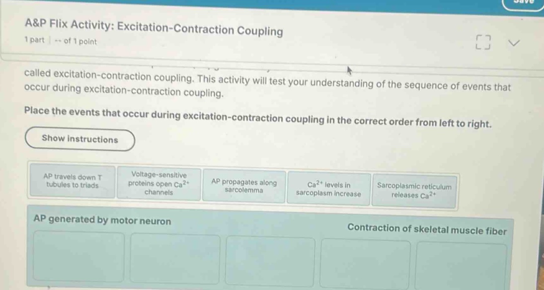
place the events that occur during excitation-contraction coupling in the correct order from left to right.
show instructions
- ap travels down t tubules to triads
- voltage-sensitive proteins open \\(\text{ca}^{2+}\\) channels
- ap propagates along sarcolemma
- \\(\text{ca}^{2+}\\) levels in sarcoplasm increase
- sarcoplasmic reticulum releases \\(\text{ca}^{2+}\\)
ap generated by motor neuron
contraction of skeletal muscle fiber
🆕 New Concept Discovered: Excitation-Contraction Coupling
How electrical signals trigger muscle contraction.
Step 1: Action potential propagation along the sarcolemma
Once an action potential (AP) is generated at the neuromuscular junction by the motor neuron, it must spread across the surface of the muscle fiber. The cell membrane of a muscle fiber is called the sarcolemma. Therefore, the first event in this sequence is the propagation of the action potential along this membrane.
- Event 1: AP propagates along sarcolemma
Step 2: Action potential traveling down the T-tubules
To trigger a contraction throughout the entire muscle cell, the electrical signal needs to reach the interior of the fiber. It does this by traveling down deep invaginations of the sarcolemma called transverse tubules (T-tubules) to reach the triads (where T-tubules meet the sarcoplasmic reticulum).
- Event 2: AP travels down T tubules to triads
Step 3: Activation of voltage-sensitive proteins
As the action potential travels down the T-tubules, the change in membrane voltage is detected by specialized voltage-sensitive proteins (DHP receptors) located in the T-tubule membrane. These proteins physically change shape in response to the electrical signal.
- Event 3: Voltage-sensitive proteins open \( \text{Ca}^{2+} \) channels
Step 4: Release of calcium from the sarcoplasmic reticulum
The conformational change of the voltage-sensitive proteins mechanically opens calcium release channels (ryanodine receptors) in the membrane of the adjacent sarcoplasmic reticulum (SR), which is the muscle cell's calcium storage site. This allows stored calcium ions to flood out into the cytosol.
- Event 4: Sarcoplasmic reticulum releases \( \text{Ca}^{2+} \)
Step 5: Increase of calcium levels in the sarcoplasm
As calcium ions exit the sarcoplasmic reticulum, they flood the cytoplasm of the muscle cell (called the sarcoplasm). This rise in intracellular calcium concentration is the final trigger that allows calcium to bind to troponin, initiating the sliding filament mechanism of muscle contraction.
- Event 5: \( \text{Ca}^{2+} \) levels in sarcoplasm increase
---

Snap & solve any problem in the app
Get step-by-step solutions on Sovi AI
Photo-based solutions with guided steps
Explore more problems and detailed explanations
The correct order of events from left to right is:
- AP propagates along sarcolemma
- AP travels down T tubules to triads
- Voltage-sensitive proteins open \( \text{Ca}^{2+} \) channels
- Sarcoplasmic reticulum releases \( \text{Ca}^{2+} \)
- \( \text{Ca}^{2+} \) levels in sarcoplasm increase