QUESTION IMAGE
Question
activity 17 : quiz
module : nervous
topic : structures / type : combination
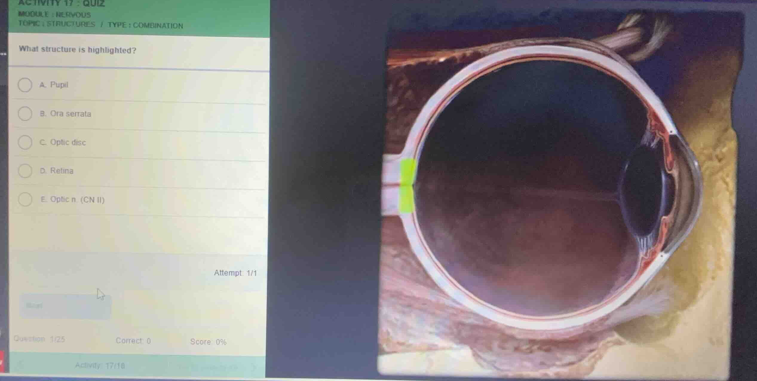
what structure is highlighted?
a. pupil
b. ora serrata
c. optic disc
d. retina
e. optic n. (cn ii)
Identify the highlighted anatomical region
Using the Eye Anatomy and Optic Nerve Anatomy knowledge points
The provided cross-sectional anatomical diagram of the eye highlights a specific region in bright green. This region is located at the posterior aspect of the eyeball, precisely where the neural retina converges and the optic nerve fibers exit the globe to form the optic nerve (CN II).
Differentiate the anatomical options
- Pupil: The anterior opening in the center of the iris.
- Ora serrata: The serrated junction between the retina and the ciliary body located anteriorly.
- Optic disc: The circular area at the back of the eye where the optic nerve connects to the retina. It is also known as the blind spot because it lacks photoreceptors. The green highlight is placed exactly at this junction on the inner wall of the posterior eye.
- Retina: The entire inner neural layer of the eye, not just this localized exit point.
- Optic n. (CN II): The nerve bundle extending posteriorly from the eyeball, whereas the highlight specifically marks the disc-like entry/exit interface on the posterior wall.

Snap & solve any problem in the app
Get step-by-step solutions on Sovi AI
Photo-based solutions with guided steps
Explore more problems and detailed explanations
- (A) Pupil
- (B) Ora serrata
- (C) Optic disc (Correct answer)
- (D) Retina
- (E) Optic n. (CN II)