QUESTION IMAGE
Question
activity 17: quiz
module: nervous
topic: structures / type: combination
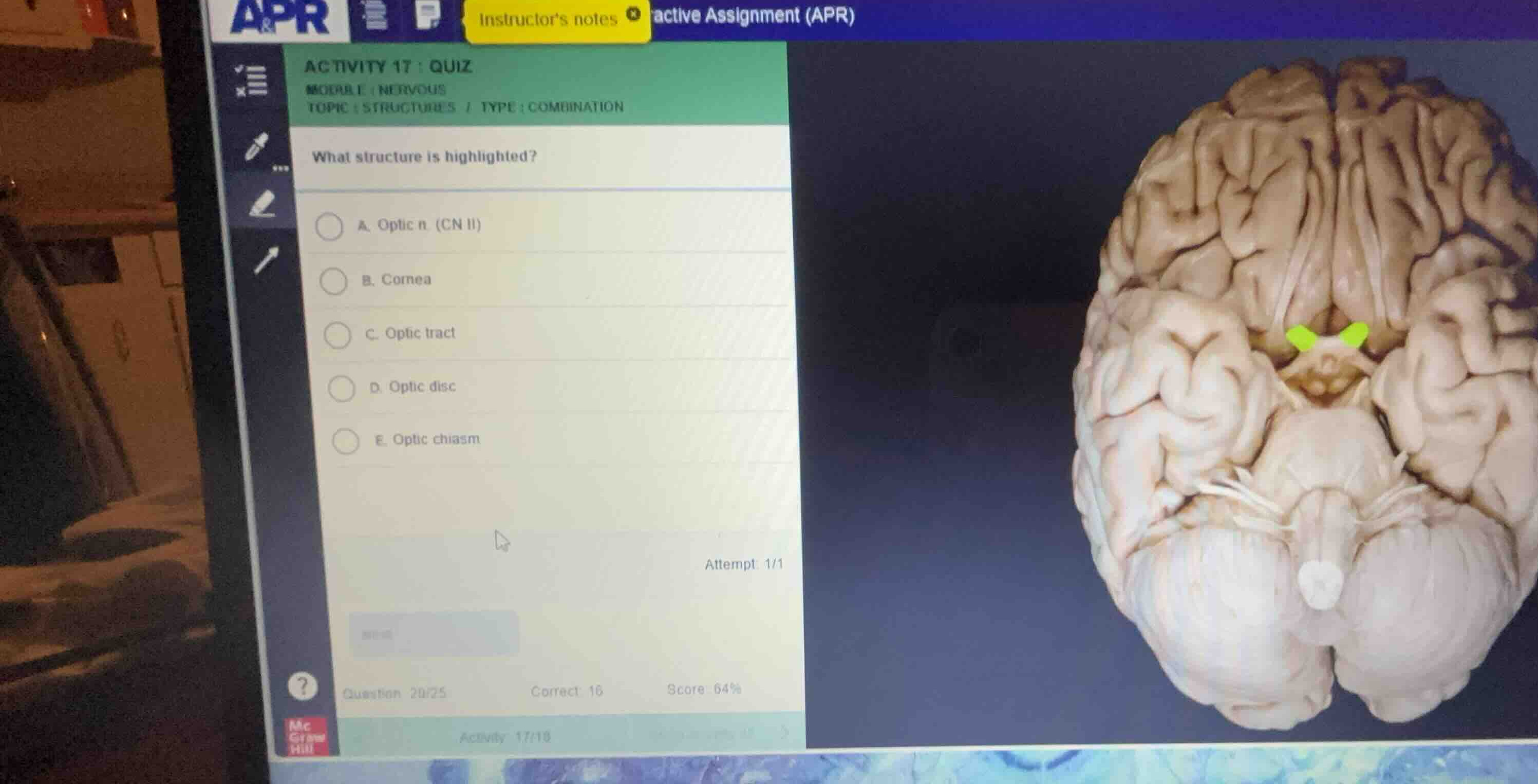
what structure is highlighted?
a. optic n. (cn ii)
b. cornea
c. optic tract
d. optic disc
e. optic chiasm
Identify the highlighted anatomical structure
Using the Optic Chiasm Anatomy knowledge point
The provided image shows an inferior (ventral) view of the human brain. The highlighted structure is the X-shaped intersection located on the ventral surface of the brain, immediately anterior to the pituitary stalk (infundibulum) and hypothalamus. This specific anatomical crossing where fibers from the nasal half of each retina decussate (cross over to the opposite side of the brain) is the optic chiasm.
Differentiate from other options
Using the Optic Chiasm Anatomy knowledge point
- Optic n. (CN II): These are the nerve bundles anterior to the chiasm, leading from the eyes.
- Optic tract: These are the fiber bundles continuing posteriorly from the chiasm toward the lateral geniculate nuclei.
- Cornea and Optic disc: These are structures located within the eyeball itself, not visible on this ventral brain specimen.
- Therefore, the highlighted X-shaped junction is the optic chiasm.

Snap & solve any problem in the app
Get step-by-step solutions on Sovi AI
Photo-based solutions with guided steps
Explore more problems and detailed explanations
- (A) Optic n. (CN II)
- (B) Cornea
- (C) Optic tract
- (D) Optic disc
- (E) Optic chiasm (Correct answer)