QUESTION IMAGE
Question
activity 10: quiz
module: cardiovascular
topic: structures / type: multiple choice (mc)
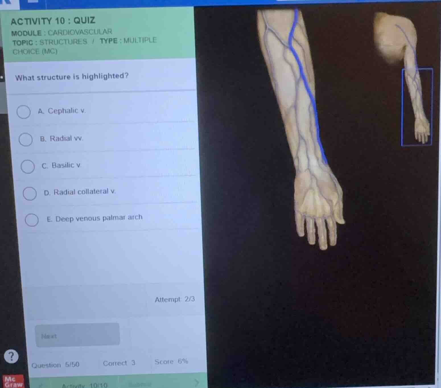
what structure is highlighted?
a. cephalic v.
b. radial vv.
c. basilic v.
d. radial collateral v.
e. deep venous palmar arch
Identify anatomical orientation
The image displays the anterior (palmar) aspect of the right forearm and hand. The thumb (lateral side) is positioned on the right side of the main anatomical model, while the pinky finger (medial side) is on the left.
Analyze the highlighted vessel
Using the Venous Anatomy knowledge point, we trace the highlighted blue vessel running along the lateral (radial) border of the forearm.
Differentiate lateral and medial superficial veins
The superficial venous system of the upper limb primarily consists of:
- The cephalic vein, which runs along the lateral (radial) side of the forearm and arm.
- The basilic vein, which runs along the medial (ulnar) side of the forearm and arm.
Since the highlighted vessel is on the lateral side (aligned with the thumb), it is the cephalic vein.
Evaluate the multiple-choice options
- Option A (Cephalic v.) matches the lateral superficial vein of the forearm.
- Option B (Radial vv.) are deep veins that typically run in pairs alongside the radial artery.
- Option C (Basilic v.) is located on the medial side of the forearm.
- Option D (Radial collateral v.) is a deeper tributary associated with the arm.
- Option E (Deep venous palmar arch) is located deep within the palm of the hand.

Snap & solve any problem in the app
Get step-by-step solutions on Sovi AI
Photo-based solutions with guided steps
Explore more problems and detailed explanations
- (A) Cephalic v. (Correct answer)
- (B) Radial vv.
- (C) Basilic v.
- (D) Radial collateral v.
- (E) Deep venous palmar arch