QUESTION IMAGE
Question
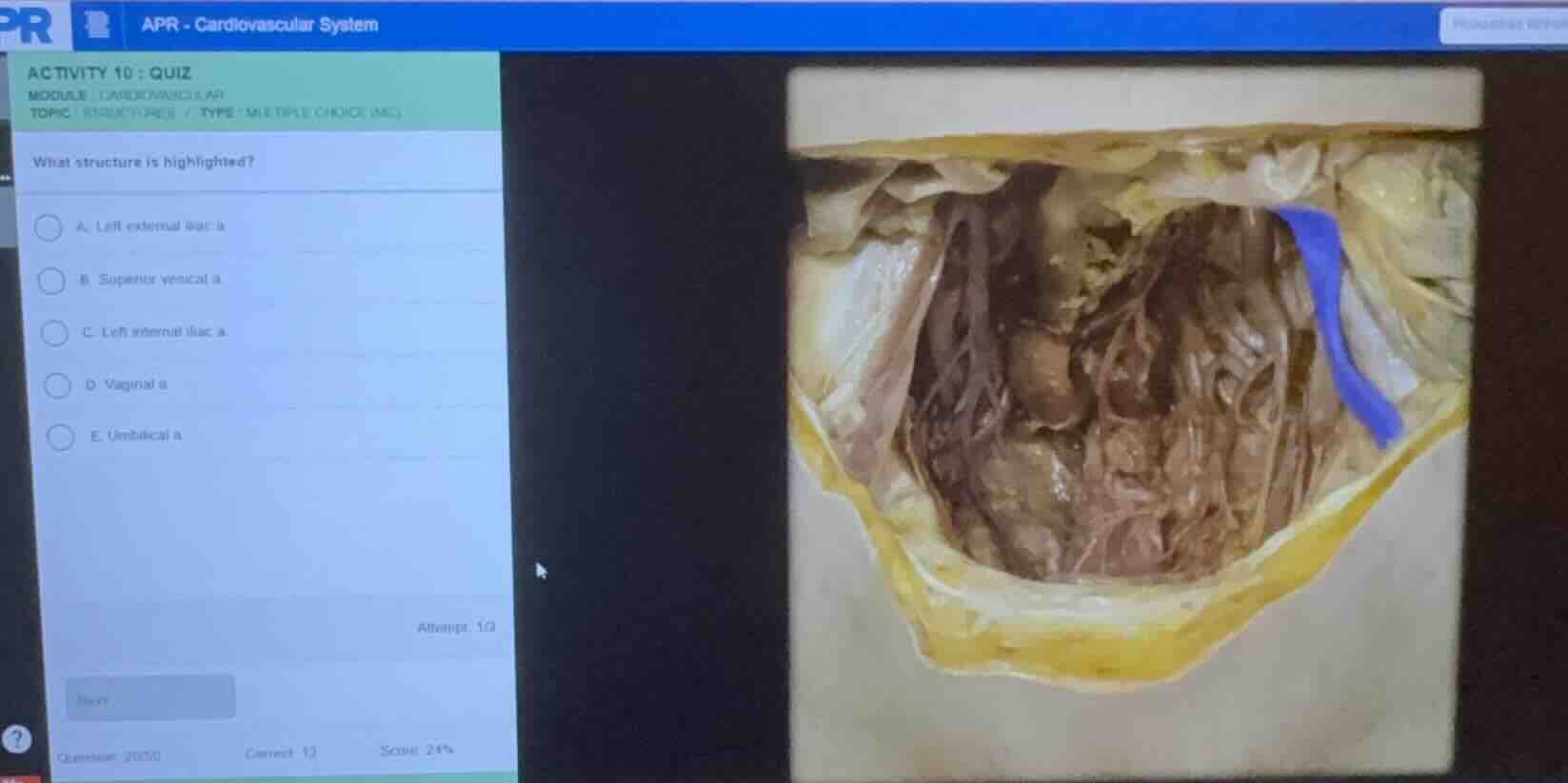
what structure is highlighted?
a. left external iliac a.
b. superior vesical a.
c. left internal iliac a.
d. vaginal a.
e. umbilical a.
Identify the anatomical region and view
The image shows a dissection of the human pelvis, viewed from an anterior/superior perspective looking down into the pelvic cavity. The anterior abdominal wall has been reflected downward. On the right side of the image (which corresponds to the anatomical left side of the body), a prominent vessel running along the lateral pelvic wall toward the anterior abdominal wall/urinary bladder is highlighted in blue.
Analyze the highlighted vessel's pathway
The highlighted vessel originates from the internal iliac artery system deep in the pelvis. It courses anteriorly and superiorly along the lateral pelvic wall, heading toward the anterior abdominal wall and the superior aspect of the urinary bladder. In a living body or complete dissection, this vessel continues anteriorly toward the umbilicus as the medial umbilical ligament (the obliterated distal portion of the umbilical artery). The patent proximal portion gives off the superior vesical arteries to supply the bladder.
Evaluate the multiple-choice options
Let's compare the anatomical pathway of the highlighted structure with the given choices:
- A. Left external iliac a.: The external iliac artery runs along the medial border of the psoas major muscle toward the inguinal ligament to become the femoral artery. The highlighted vessel is medial to this path and courses anteriorly toward the anterior abdominal wall/bladder.
- B. Superior vesical a.: These are branches that typically arise from the patent proximal part of the umbilical artery to supply the upper part of the bladder. While related, the highlighted structure is the main trunk continuing anteriorly.
- C. Left internal iliac a.: The main internal iliac artery is a short, thick trunk located deeper and more posteriorly in the pelvis before it divides into anterior and posterior divisions.
- D. Vaginal a.: This is a branch of the internal iliac artery in females that runs medially and inferiorly to supply the vagina, which does not match this long anteriorly-coursing vessel.
- E. Umbilical a.: The umbilical artery arises from the anterior division of the internal iliac artery. Its patent proximal part gives off superior vesical branches, while its distal part obliterates to form the medial umbilical ligament running up the anterior abdominal wall. The highlighted blue structure represents this prominent vessel.
Confirm the correct anatomical identification
The highlighted structure is the left umbilical artery (specifically its patent proximal portion and its continuation toward the anterior wall), which is a major branch of the anterior division of the internal iliac artery.

Snap & solve any problem in the app
Get step-by-step solutions on Sovi AI
Photo-based solutions with guided steps
Explore more problems and detailed explanations
- (A) Left external iliac a.
- (B) Superior vesical a.
- (C) Left internal iliac a.
- (D) Vaginal a.
- (E) Umbilical a. (Correct answer)