QUESTION IMAGE
Question
for review
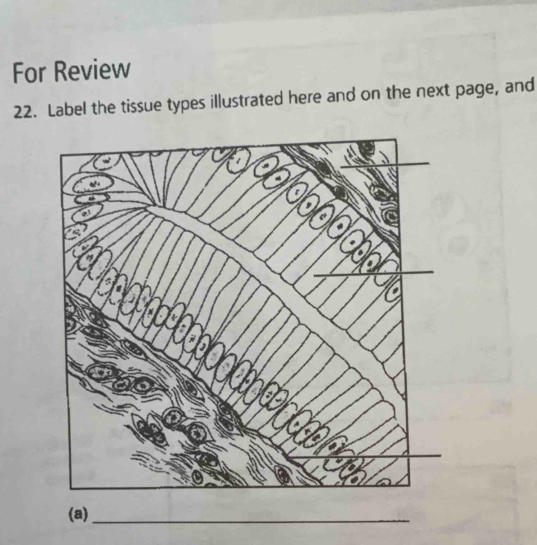
- label the tissue types illustrated here and on the next page, and
(a)
🆕 New Concept Discovered: Epithelial Tissue Classification
Identifying tissues by cell shape and layers.
Step 1: Analyze the overall tissue structure (a)
To identify the main tissue type shown in diagram (a), we look at the organization of the cells.
- There is a prominent layer of tall, column-like cells lined up side-by-side.
- These cells have elongated nuclei located near the base of the cells.
- This single layer of tall cells is characteristic of simple columnar epithelium.
Step 2: Identify the specific labeled structures
Let's identify the three pointer lines from top to bottom:
- Top pointer: Points to the underlying connective tissue layer beneath the basement membrane (specifically, the connective tissue or lamina propria containing fibroblasts and extracellular fibers).
- Middle pointer: Points directly to a single, tall column-shaped cell, which is a simple columnar epithelial cell (or its nucleus / cytoplasm).
- Bottom pointer: Points to the thin boundary line at the base of the epithelial cells, which is the basement membrane.

Snap & solve any problem in the app
Get step-by-step solutions on Sovi AI
Photo-based solutions with guided steps
Explore more problems and detailed explanations
- Overall Tissue Type (a): Simple columnar epithelium
- Top label: Connective tissue (or Lamina propria)
- Middle label: Simple columnar epithelial cell (or Nucleus of columnar cell)
- Bottom label: Basement membrane