QUESTION IMAGE
Question
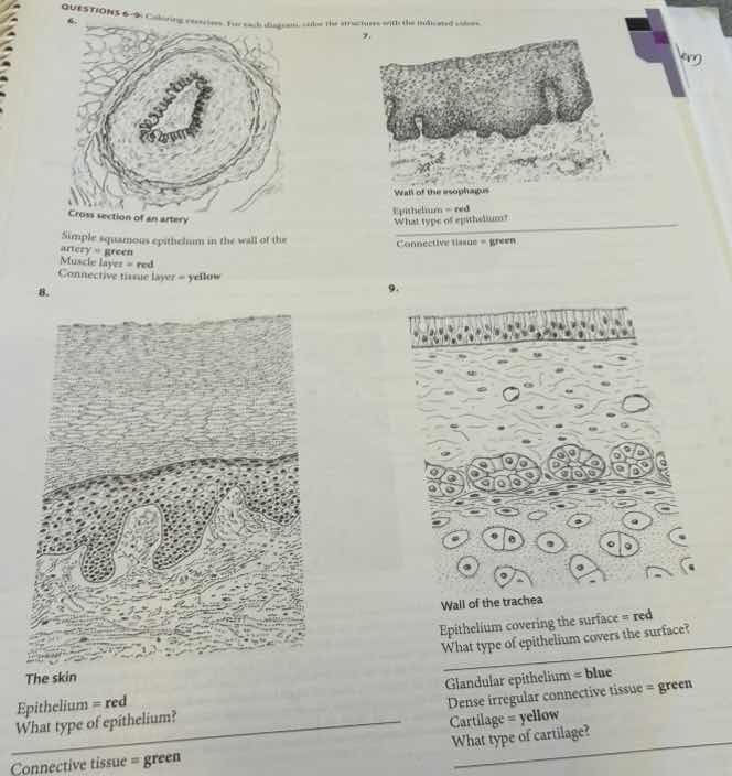
questions 6-9: coloring exercises. for each diagram, color the structures with the indicated colors.
6.
cross section of an artery
simple squamous epithelium in the wall of the artery = green
muscle layer = red
connective tissue layer = yellow
7.
wall of the esophagus
epithelium = red
what type of epithelium?
connective tissue = green
8.
the skin
epithelium = red
what type of epithelium?
connective tissue = green
9.
wall of the trachea
epithelium covering the surface = red
what type of epithelium covers the surface?
glandular epithelium = blue
dense irregular connective tissue = green
cartilage = yellow
what type of cartilage?
🆕 New Concept Discovered: Epithelial and Connective Tissue Classification
Identifying tissue types by cell shape, layering, and extracellular matrix.
Step 1: Analyze Question 7 (Wall of the esophagus)
The esophagus is a muscular tube that transports food from the mouth to the stomach. Because it experiences significant friction and wear from swallowed food, it requires a protective barrier.
- Epithelium type: The image shows multiple layers of cells (stratified) where the surface cells are flattened (squamous). Since it is a moist internal lining, it is non-keratinized.
- Classification: Stratified squamous epithelium (non-keratinized).
Step 2: Analyze Question 8 (The skin)
The skin (epidermis) forms the outermost protective barrier of the body, facing constant abrasion and water loss.
- Epithelium type: It consists of many layers of cells (stratified) with flattened cells at the surface (squamous). To prevent water loss and provide extra protection, the outer layers are filled with a tough, dead protein called keratin.
- Classification: Keratinized stratified squamous epithelium.
Step 3: Analyze Question 9 (Wall of the trachea)
The trachea (windpipe) conducts air to the lungs and needs to trap and sweep away inhaled dust and debris.
- Epithelium type: It appears to have multiple layers because the cell nuclei are at different heights, but every cell actually touches the basement membrane (pseudostratified). The cells are tall and column-like (columnar) and have hair-like projections (cilia) on their free surface to move mucus.
- Classification: Pseudostratified ciliated columnar epithelium.
- Cartilage type: The trachea is kept open by stiff, supportive rings made of the most common, glassy-looking cartilage in the body, which contains fine collagen fibers.
- Classification: Hyaline cartilage.

Snap & solve any problem in the app
Get step-by-step solutions on Sovi AI
Photo-based solutions with guided steps
Explore more problems and detailed explanations
- Question 7 (Wall of the esophagus): Stratified squamous epithelium (non-keratinized)
- Question 8 (The skin): Keratinized stratified squamous epithelium
- Question 9 (Wall of the trachea):
- Type of epithelium: Pseudostratified ciliated columnar epithelium
- Type of cartilage: Hyaline cartilage