QUESTION IMAGE
Question
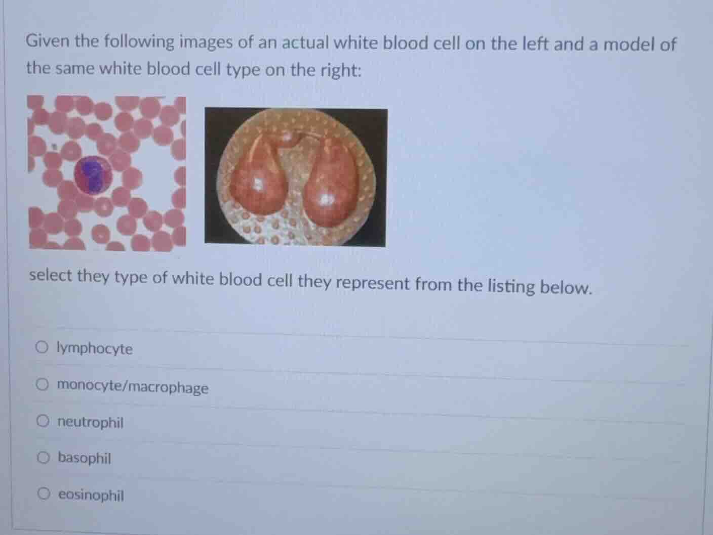
given the following images of an actual white blood cell on the left and a model of the same white blood cell type on the right:
select they type of white blood cell they represent from the listing below.
lymphocyte
monocyte/macrophage
neutrophil
basophil
eosinophil
Identify cell characteristics
Using the White Blood Cells knowledge point:
- The micrograph on the left shows a white blood cell with a distinct bilobed nucleus and cytoplasm filled with bright reddish-orange granules.
- The model on the right depicts a bilobed nucleus (two large lobes connected by a thin strand) and a cytoplasm packed with prominent granules.
Match with leukocyte types
- Eosinophils are characterized by a bilobed nucleus and prominent, reddish-orange (eosinophilic) granules in their cytoplasm.
- Lymphocytes have a large, round nucleus with very little cytoplasm and no prominent granules.
- Monocytes are large agranulocytes with a kidney- or horseshoe-shaped nucleus.
- Neutrophils typically have multi-lobed nuclei (3 to 5 lobes) with pale, neutral-staining granules.
- Basophils have a bilobed or bent nucleus that is usually obscured by dark blue-purple, coarse granules.
Select the correct option
The visual evidence of a bilobed nucleus combined with bright reddish-orange granules uniquely identifies this cell as an eosinophil.

Snap & solve any problem in the app
Get step-by-step solutions on Sovi AI
Photo-based solutions with guided steps
Explore more problems and detailed explanations
- lymphocyte
- monocyte/macrophage
- neutrophil
- basophil
- eosinophil (Correct answer)