QUESTION IMAGE
Question
complete the labeling activity.
place the appropriate labels in their respective targets.
show instructions
goblet cells
simple cuboidal
stratified squamous
simple squamous
pseudostratified columnar
basement membrane
cilia
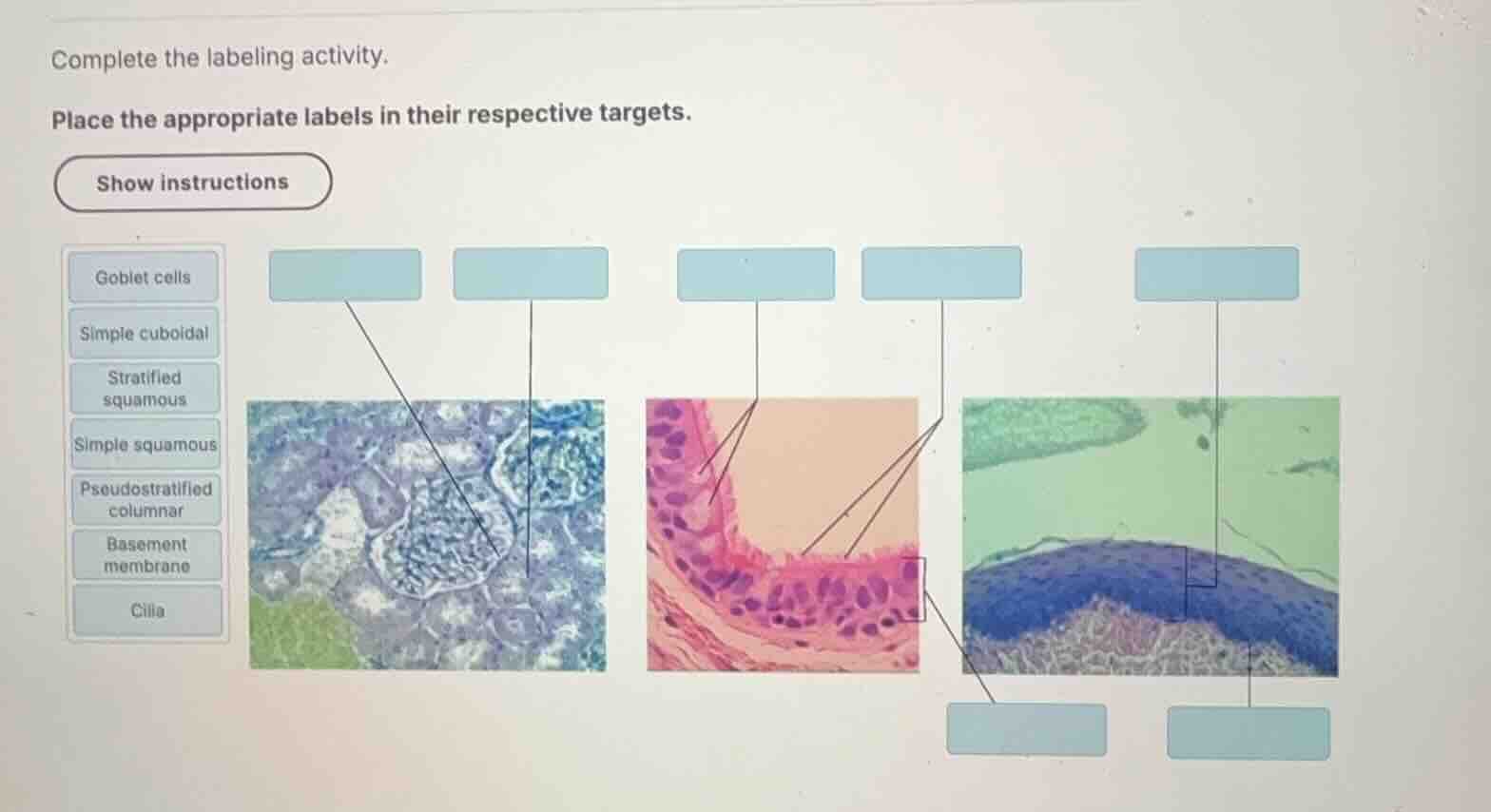
Identify the first micrograph (left)
The first image shows kidney tubules and a glomerulus. The pointer on the left points to the single layer of cube-shaped cells lining a tubule, which is Simple cuboidal epithelium. The pointer on the right points to the thin, flat cells forming the outer capsule (Bowman's capsule) or the capillary wall, which represents Simple squamous epithelium. This aligns with Simple Cuboidal Epithelium and Simple Squamous Epithelium Locations.
Identify the second micrograph (middle)
The middle image shows a respiratory lining. The tissue is Pseudostratified columnar epithelium, characterized by nuclei at different levels. The pointers point to specific structures:
- The upper pointer points to the mucus-secreting Goblet cells.
- The lower pointer points to the hair-like projections on the apical surface, which are Cilia.
- The bracket on the right side of this tissue points to the entire epithelial layer, which is Pseudostratified columnar.
Identify the third micrograph (right)
The third image shows a thick, multi-layered epithelium with flattened cells at the apical surface, characteristic of Stratified squamous epithelium.
- The top pointer points to the thick epithelial layer itself, which is Stratified squamous.
- The bottom pointer points to the boundary line separating the epithelium from the underlying connective tissue, which is the Basement membrane.

Snap & solve any problem in the app
Get step-by-step solutions on Sovi AI
Photo-based solutions with guided steps
Explore more problems and detailed explanations
| Target Location | Correct Label |
|---|---|
| First Micrograph (Left) - Right Pointer | Simple squamous |
| Second Micrograph (Middle) - Upper Pointer | Goblet cells |
| Second Micrograph (Middle) - Lower Pointer | Cilia |
| Second Micrograph (Middle) - Right Bracket | Pseudostratified columnar |
| Third Micrograph (Right) - Top Pointer | Stratified squamous |
| Third Micrograph (Right) - Bottom Pointer | Basement membrane |