QUESTION IMAGE
Question
activity 10: quiz
module: cardiovascular
topic: structures / type: multiple choice (mc)
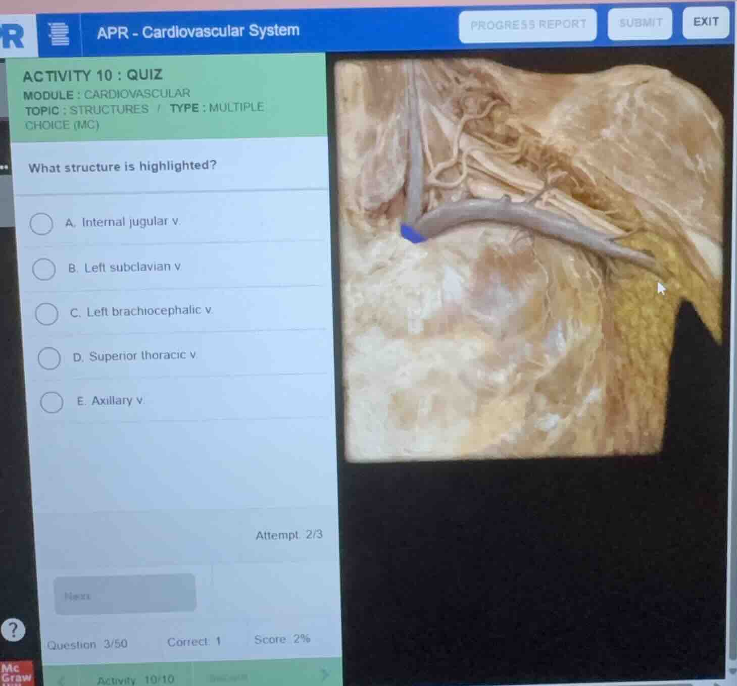
what structure is highlighted?
a. internal jugular v.
b. left subclavian v.
c. left brachiocephalic v.
d. superior thoracic v.
e. axillary v.
Identify the anatomical region
The image shows a cadaveric dissection of the upper thoracic and lower neck region on the left side of the body. We can observe major venous structures returning blood toward the heart.
Trace the major veins
We can identify a large horizontal vein running from the left upper limb/shoulder area toward the midline.
- The vein coming from the arm/axilla is the axillary vein, which becomes the subclavian vein as it crosses the first rib.
- A vertical vein descending from the neck is the internal jugular vein.
- The junction where the internal jugular vein and the subclavian vein unite forms the brachiocephalic vein.
Analyze the highlighted structure
The blue highlighted region is located at the medial-most portion of this venous confluence, just as the left subclavian vein and left internal jugular vein merge to form the left brachiocephalic vein. Specifically, the highlight is placed at the very beginning of the left brachiocephalic vein (or the junction forming it), directing blood medially toward the superior vena cava.
Evaluate the multiple-choice options
- A. Internal jugular v.: This is the vertical vein descending from the neck, which is visible but not highlighted in blue.
- B. Left subclavian v.: This is the long horizontal vein segment lateral to the junction, which is grey/unhighlighted.
- C. Left brachiocephalic v.: This is the medial continuation formed by the union of the subclavian and internal jugular veins, which matches the highlighted blue region.
- D. Superior thoracic v.: This is a much smaller tributary, not the major highlighted trunk.
- E. Axillary v.: This lies more laterally, deep to the shoulder region.
Therefore, the highlighted structure is the left brachiocephalic vein.

Snap & solve any problem in the app
Get step-by-step solutions on Sovi AI
Photo-based solutions with guided steps
Explore more problems and detailed explanations
- (A) Internal jugular v.
- (B) Left subclavian v.
- (C) Left brachiocephalic v. (Correct answer)
- (D) Superior thoracic v.
- (E) Axillary v.