QUESTION IMAGE
Question
students instructions
introduction
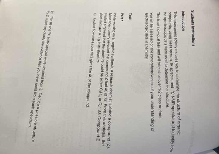
this assessment activity requires you to determine the structure of organic compounds, using mass spectra, ir spectra, and \\(^{13}\text{c}\\) nmr spectra and to justify how the spectroscopic data were used to determine the structure.
this is an individual task and will take place over 1-2 class periods.
you will be assessed on the comprehensiveness of your understanding of spectroscopic data in chemistry.
task
part 1
while working on an organic synthesis, a research chemist isolated a compound (z). mass spectrometry revealed that compound z had \\(m_r\\) of 72. from this analysis, the student proposed that the structure could be either \\(\text{c}_5\text{h}_{12}\\) or \\(\text{c}_4\text{h}_8\text{o}\\). compound z does not have a ring in its structure.
a) explain how mass spec data gives the \\(m_r\\) of the compound.
b) the ir and \\(^{13}\text{c}\\) nmr spectra were obtained from z. deduce a possible structure for z indicating clearly the evidence that you have used from each spectrum.
Explain mass spec determination of molecular mass
Mass spectrometry determines the molecular mass \(M_r\) of a compound by ionizing the molecules and measuring their mass-to-charge ratio (\(m/z\)). The intact molecule loses one electron to form a radical cation, known as the molecular ion (\(M^{\bullet+}\)). The peak with the highest \(m/z\) value in the simplified spectrum (excluding small isotope peaks like \(M+1\)) corresponds to this molecular ion, directly revealing the molecular mass of the compound.
Analyze the missing spectral data for structure deduction
To deduce a possible structure for compound Z, the actual Infrared (IR) and Carbon-13 (\(^{13}\text{C}\)) NMR spectra are required. Since these spectra are not visually provided in the image, we can outline the analytical method:
- IR Spectroscopy: Identifies functional groups by characteristic bond vibration frequencies (e.g., \(\text{C=O}\) stretch around \(1700\text{ cm}^{-1}\), \(\text{O-H}\) stretch around \(3300\text{ cm}^{-1}\)).
- \(^{13}\text{C}\) NMR Spectroscopy: Determines the number of unique carbon environments and their chemical shifts, indicating the carbon skeleton symmetry and neighboring atoms.

Snap & solve any problem in the app
Get step-by-step solutions on Sovi AI
Photo-based solutions with guided steps
Explore more problems and detailed explanations
Question a
Mass spectrometry determines the molecular mass (\(M_r\)) of a compound by ionizing the sample to produce a molecular ion, \(M^{\bullet+}\). The mass-to-charge ratio (\(m/z\)) of the molecular ion peak (the peak furthest to the right in the main spectrum, representing the unbroken ionized molecule) corresponds directly to the molecular mass of the compound.
Question b
To deduce the structure of compound Z, the corresponding IR and \(^{13}\text{C}\) NMR spectra must be analyzed:
- IR Spectrum: Look for key absorption bands to identify functional groups. For example, a strong peak near \(1700\text{ cm}^{-1}\) indicates a carbonyl group (\(\text{C=O}\)), which would confirm the formula \(\text{C}_4\text{H}_8\text{O}\) rather than \(\text{C}_5\text{H}_{12}\).
- \(^{13}\text{C}\) NMR Spectrum: Count the number of peaks to determine the number of unique carbon environments, and use chemical shifts to identify the types of carbon atoms present to assemble the final carbon skeleton.