QUESTION IMAGE
Question
i
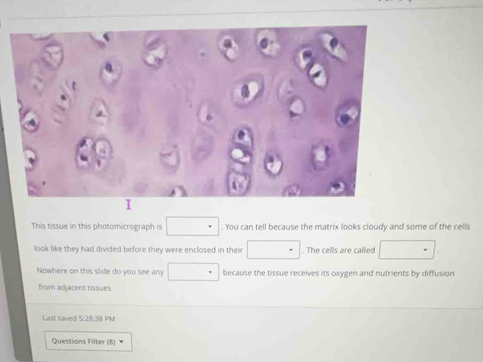
this tissue in this photomicrograph is dropdown. you can tell because the matrix looks cloudy and some of the cells look like they had divided before they were enclosed in their dropdown. the cells are called dropdown.
nowhere on this slide do you see any dropdown because the tissue receives its oxygen and nutrients by diffusion from adjacent tissues.
🆕 New Concept Discovered: Cartilage Histology
Identifying specialized connective tissues and their structural features.
Step 1: Identify the tissue type
Looking at the photomicrograph, we see cells residing in small, clear spaces (lacunae) scattered throughout a smooth, glassy, or slightly cloudy extracellular matrix. Some cells are grouped together in pairs or small clusters (isogenous groups), which indicates they recently divided before being trapped in the matrix. This appearance is characteristic of hyaline cartilage.
Step 2: Identify the cell compartments
The small cavities or chambers in the extracellular matrix that enclose these cartilage cells are called lacunae (singular: lacuna).
Step 3: Identify the cell type
The mature cells found within the lacunae of cartilage are called chondrocytes.
Step 4: Identify the missing vascular structures
Cartilage is an avascular tissue, meaning it does not contain its own blood supply. It relies entirely on diffusion from the surrounding connective tissue (the perichondrium) for oxygen and nutrients. Therefore, you do not see any blood vessels on this slide.

Snap & solve any problem in the app
Get step-by-step solutions on Sovi AI
Photo-based solutions with guided steps
Explore more problems and detailed explanations
- First blank: hyaline cartilage
- Second blank: lacunae
- Third blank: chondrocytes
- Fourth blank: blood vessels