QUESTION IMAGE
Question
proximal epiphysis
metaphysis
diaphysis
metaphysis
distal epiphysis
spongy bone
epiphyseal line
red bone marrow
endosteum
compact bone
medullary cavity
yellow bone marrow
periosteum
nutrient artery
articular cartilage
🆕 New Concept Discovered: Anatomy of a Long Bone
Understanding the structural regions and tissues of long bones.
Step 1: Identify the highlighted structure
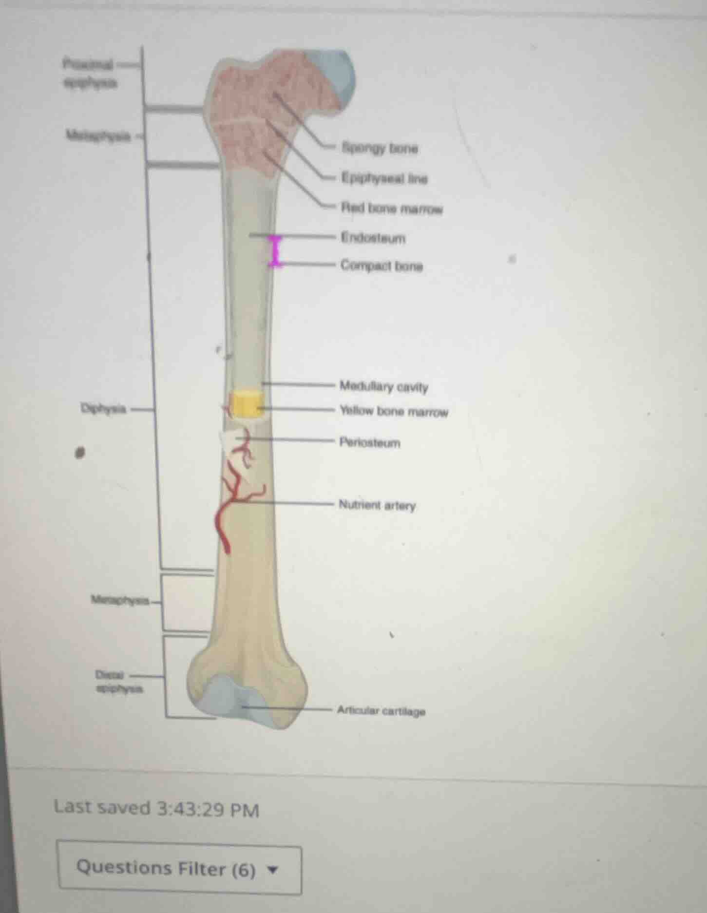
The image shows a diagram of a long bone (specifically a femur) with various anatomical parts labeled.
There is a purple bracket with a capital letter I next to it on the upper-middle shaft of the bone.
Step 2: Trace the bracket lines
Looking closely at the purple bracket labeled I:
- The top horizontal line of the bracket aligns with the label Endosteum.
- The bottom horizontal line of the bracket aligns with the label Compact bone.
The bracket spans the thickness of the hard, outer wall of the bone shaft (diaphysis).
Step 3: Determine the anatomical layer
- Compact bone is the dense, hard outer layer of bone tissue that provides strength and protection.
- Endosteum is the thin vascular membrane of connective tissue that lines the inner surface of the bony tissue (the medullary cavity).
- Periosteum is the membrane that covers the outer surface of all bones.
The bracket I specifically highlights the thick, dense layer of the bone wall, which is the Compact bone (and its inner lining, the Endosteum). Depending on the specific multiple-choice question associated with this standard diagram, the bracket I is pointing to the Compact bone layer of the diaphysis.

Snap & solve any problem in the app
Get step-by-step solutions on Sovi AI
Photo-based solutions with guided steps
Explore more problems and detailed explanations
The bracket labeled I indicates the Compact bone (specifically highlighting the thickness of the compact bone layer of the diaphysis, lined internally by the Endosteum).