QUESTION IMAGE
Question
place the appropriate labels in their respective targets.
show instructions
stratified squamous keratinized epithelium
simple epithelia
stratified columnar epithelium
stratified cuboidal epithelium
simple columnar epithelium
pseudostratified columnar epithelium
simple squamous epithelium
simple cuboidal epithelium
transitional epithelium
stratified squamous nonkeratinized epithelium
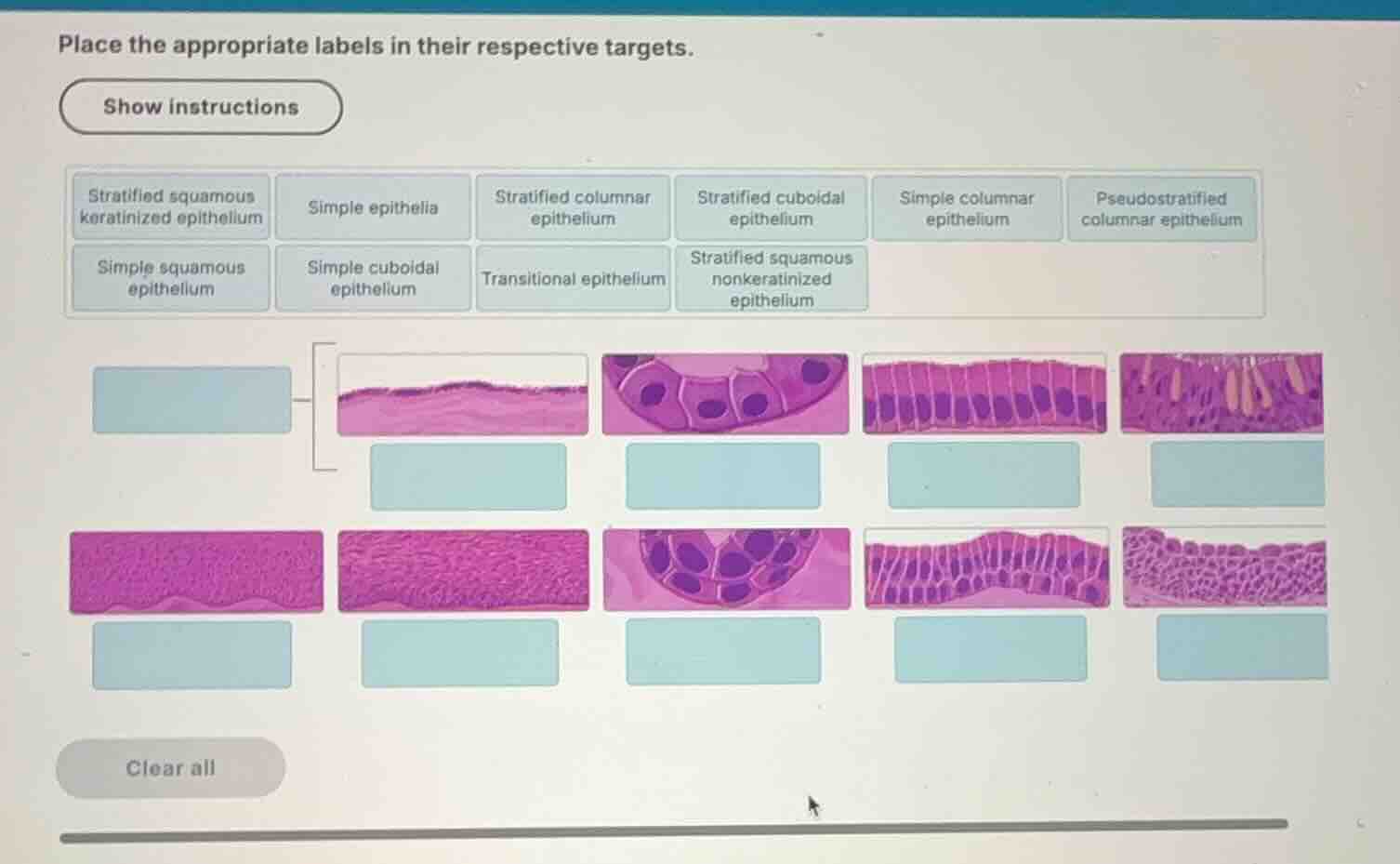
Classify the grouping label
The bracket on the left groups the first two histological diagrams in the top row. Looking at these two diagrams, they represent simple squamous epithelium (a single layer of flat cells) and simple cuboidal epithelium (a single layer of cube-shaped cells). Therefore, the grouping label on the far left is Simple epithelia.
Identify top row tissues
- First diagram (top left, grouped): Shows a single layer of extremely flat, thin cells resting on a basement membrane. This is Simple squamous epithelium.
- Second diagram (top, second from left): Shows a single layer of cube-shaped cells with central, spherical nuclei forming a duct-like ring. This is Simple cuboidal epithelium, which is a concept you have already mastered (Simple Cuboidal Epithelium).
- Third diagram (top, third from left): Shows a single layer of tall, rectangular cells with oval nuclei aligned near the basal region. This is Simple columnar epithelium.
- Fourth diagram (top right): Shows a single layer of columnar cells of varying heights, where all cells contact the basement membrane but not all reach the apical surface, giving a layered appearance. Cilia are visible on the apical surface. This is Pseudostratified columnar epithelium.
Identify bottom row tissues
- First diagram (bottom left): Shows multiple layers of cells, with the apical layers being flat and dead (keratinized, forming a thick, uniform pink layer on top). This is Stratified squamous keratinized epithelium.
- Second diagram (bottom, second from left): Shows multiple layers of cells, with flat apical cells that retain their nuclei (nonkeratinized). This is Stratified squamous nonkeratinized epithelium.
- Third diagram (bottom, third from left): Shows two layers of cube-shaped cells forming a duct. This is Stratified cuboidal epithelium.
- Fourth diagram (bottom, fourth from left): Shows multiple layers of tall, columnar cells. This is Stratified columnar epithelium.
- Fifth diagram (bottom right): Shows multiple layers of cells that can change shape (dome-shaped or scalloped apical cells when relaxed). This is Transitional epithelium.

Snap & solve any problem in the app
Get step-by-step solutions on Sovi AI
Photo-based solutions with guided steps
Explore more problems and detailed explanations
The correct labels for each target box in the diagram are:
- Left Grouping Bracket (top row, first two images):
Simple epithelia
- Top Row (from left to right):
Simple squamous epitheliumSimple cuboidal epitheliumSimple columnar epitheliumPseudostratified columnar epithelium
- Bottom Row (from left to right):
Stratified squamous keratinized epitheliumStratified squamous nonkeratinized epitheliumStratified cuboidal epitheliumStratified columnar epitheliumTransitional epithelium