QUESTION IMAGE
Question
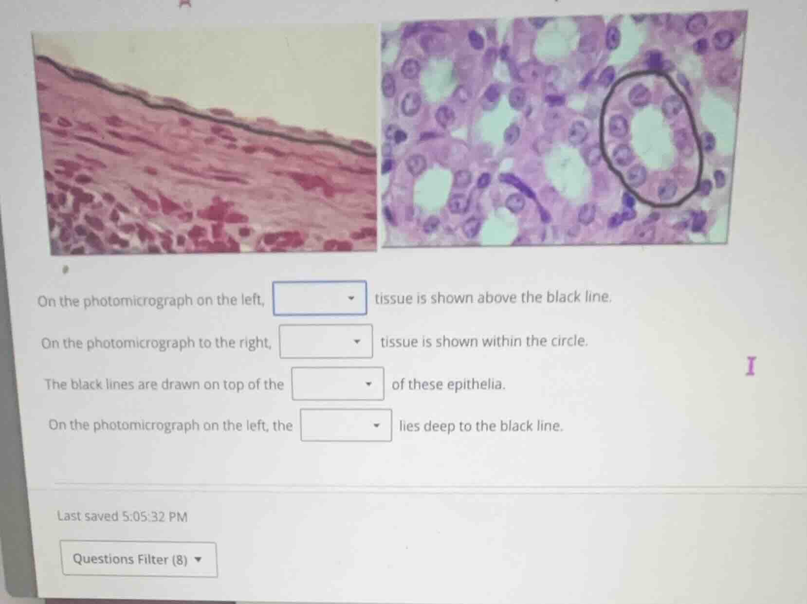
on the photomicrograph on the left, dropdown tissue is shown above the black line.
on the photomicrograph to the right, dropdown tissue is shown within the circle.
the black lines are drawn on top of the dropdown of these epithelia.
on the photomicrograph on the left, the dropdown lies deep to the black line.
🆕 New Concept Discovered: Epithelial Tissue Classification
Identifying tissues by cell shape and layers.
Step 1: Identify the tissue on the left
Looking at the left photomicrograph, we see a single layer of highly flattened, scale-like cells resting on a thin boundary.
- Cell shape: Extremely flat (squamous), with flat, disc-shaped nuclei.
- Layer count: A single layer (simple).
Therefore, the tissue shown above the black line is simple squamous epithelial tissue.
Step 2: Identify the tissue on the right
Looking at the right photomicrograph, we see a ring of cells forming a duct or tubule (circled in black).
- Cell shape: Cube-shaped (cuboidal) with round, centrally located nuclei.
- Layer count: A single layer forming a ring around a central space (lumen).
Therefore, the tissue shown within the circle is simple cuboidal epithelial tissue.
Step 3: Identify the boundary marked by the black lines
In both images, the black lines (the line on the left and the circle on the right) trace the boundary that anchors the epithelial cells to the underlying connective tissue.
- This thin, non-cellular adhesive sheet is called the basement membrane.
Step 4: Identify what lies deep to the boundary on the left
Epithelial tissues are always supported by an underlying layer of connective tissue.
- On the left image, the tissue lying deep to (below) the black line (basement membrane) is connective tissue (specifically, loose connective tissue/lamina propria).

Snap & solve any problem in the app
Get step-by-step solutions on Sovi AI
Photo-based solutions with guided steps
Explore more problems and detailed explanations
- On the photomicrograph on the left, simple squamous tissue is shown above the black line.
- On the photomicrograph to the right, simple cuboidal tissue is shown within the circle.
- The black lines are drawn on top of the basement membrane of these epithelia.
- On the photomicrograph on the left, the connective tissue lies deep to the black line.