QUESTION IMAGE
Question
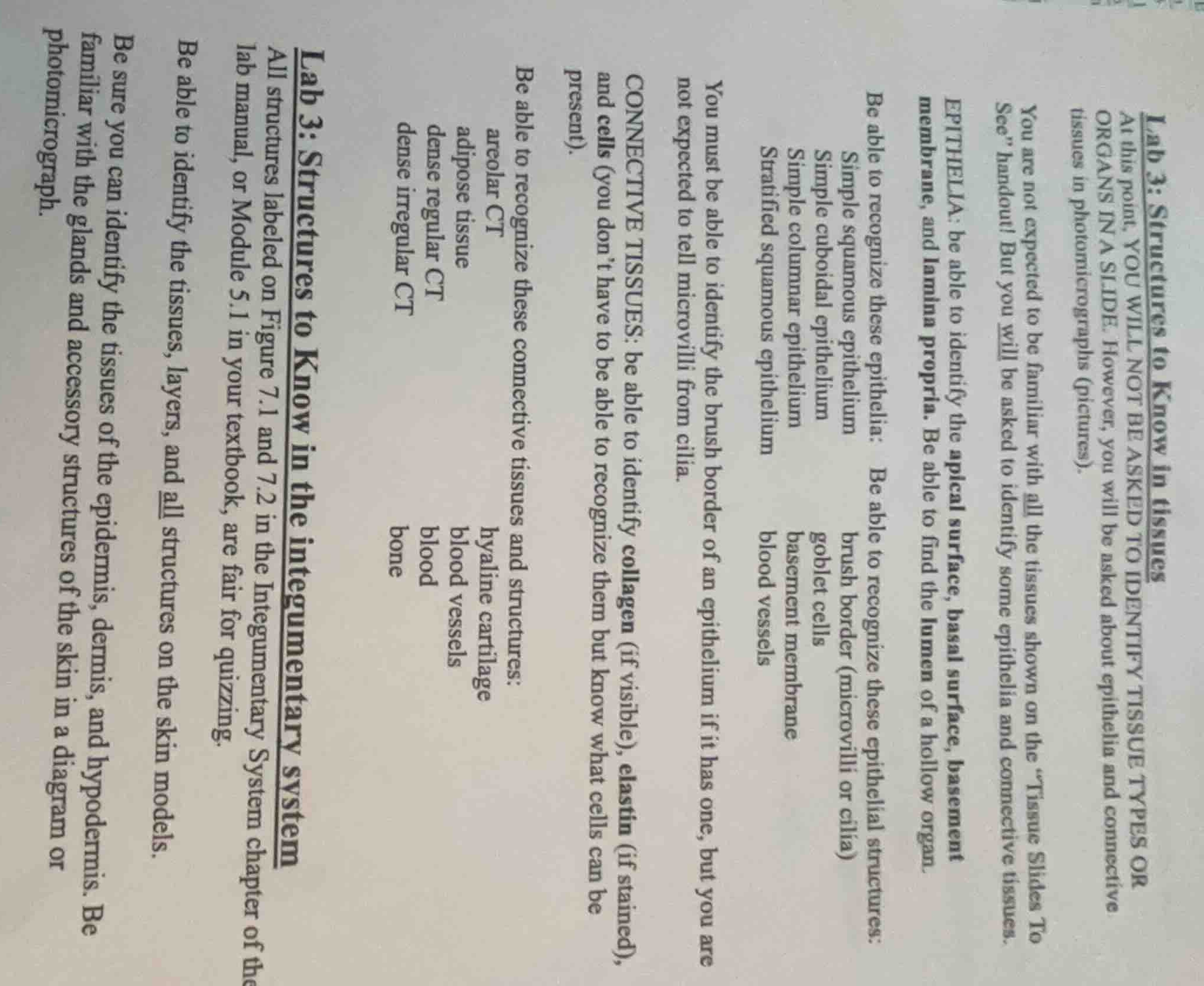
lab 3: structures to know in tissues
at this point, you will not be asked to identify tissue types or organs in a slide. however, you will be asked about epithelia and connective tissues in photomicrographs (pictures).
you are not expected to be familiar with all the tissues shown on the \tissue slides to see\ handout! but you will be asked to identify some epithelia and connective tissues.
epithelia: be able to identify the apical surface, basal surface, basement membrane, and lamina propria. be able to find the lumen of a hollow organ.
be able to recognize these epithelia:
simple squamous epithelium
simple cuboidal epithelium
simple columnar epithelium
stratified squamous epithelium
be able to recognize these epithelial structures:
brush border (microvilli or cilia)
goblet cells
basement membrane
blood vessels
you must be able to identify the brush border of an epithelium if it has one, but you are not expected to tell microvilli from cilia.
connective tissues: be able to identify collagen (if visible), elastin (if stained), and cells (you dont have to be able to recognize them but know what cells can be present).
be able to recognize these connective tissues and structures:
areolar ct
adipose tissue
dense regular ct
dense irregular ct
hyaline cartilage
blood vessels
blood
bone
lab 3: structures to know in the integumentary system
all structures labeled on figure 7.1 and 7.2 in the integumentary system chapter of the lab manual, or module 5.1 in your textbook, are fair for quizzing.
be able to identify the tissues, layers, and all structures on the skin models.
be sure you can identify the tissues of the epidermis, dermis, and hypodermis. be familiar with the glands and accessory structures of the skin in a diagram or photomicrograph.
🆕 New Concept Discovered: Histological Identification
Recognizing tissue types and structures under a microscope.
Step 1: Transcribe Epithelial Tissue Requirements
The document outlines the specific epithelial tissues and associated structures you need to recognize:
- Epithelia Types:
- Simple squamous epithelium
- Simple cuboidal epithelium
- Simple columnar epithelium
- Stratified squamous epithelium
- Epithelial Structures:
- Apical surface, basal surface, basement membrane, and lamina propria
- Lumen of a hollow organ
- Brush border (microvilli or cilia) — Note: You must identify if a brush border is present, but you are not expected to distinguish microvilli from cilia.
- Goblet cells
- Basement membrane
- Blood vessels
Step 2: Transcribe Connective Tissue Requirements
The document lists the connective tissues, fibers, and structures you must identify:
- Fibers and Cells:
- Collagen (if visible)
- Elastin (if stained)
- Cells (you do not need to identify specific cell types, but you must know which cells can be present in these tissues)
- Connective Tissue (CT) Types:
- Areolar CT
- Adipose tissue
- Dense regular CT
- Dense irregular CT
- Hyaline cartilage
- Blood vessels
- Blood
- Bone
Step 3: Transcribe Integumentary System Requirements
The final section details the structures of the skin you need to know:
- Layers: Epidermis, dermis, and hypodermis.
- Anatomical Models: All structures labeled on Figure 7.1 and 7.2 in the Integumentary System chapter of your lab manual (or Module 5.1 in your textbook).
- Microscopic Structures: Glands and accessory structures of the skin in both diagrams and photomicrographs.

Snap & solve any problem in the app
Get step-by-step solutions on Sovi AI
Photo-based solutions with guided steps
Explore more problems and detailed explanations
Based on your lab study guide, here is the checklist of what you need to study for your quiz:
1. Epithelial Tissues & Structures
- Identify these types: Simple squamous, simple cuboidal, simple columnar, and stratified squamous.
- Identify these features: Apical surface, basal surface, basement membrane, lamina propria, and the lumen of hollow organs.
- Identify these specialized structures: Brush border (presence only), goblet cells, basement membrane, and nearby blood vessels.
2. Connective Tissues & Structures
- Identify these types: Areolar CT, adipose tissue, dense regular CT, dense irregular CT, hyaline cartilage, blood, and bone.
- Identify these components: Collagen fibers (if visible), elastin fibers (if stained), general cells, and blood vessels.
3. Integumentary System (Skin)
- Identify these layers: Epidermis, dermis, and hypodermis.
- Identify on models & diagrams: All structures labeled in Figures 7.1 and 7.2 of your lab manual (or Module 5.1 in your textbook).
- Identify under the microscope: Glands and accessory structures of the skin in photomicrographs.