QUESTION IMAGE
Question
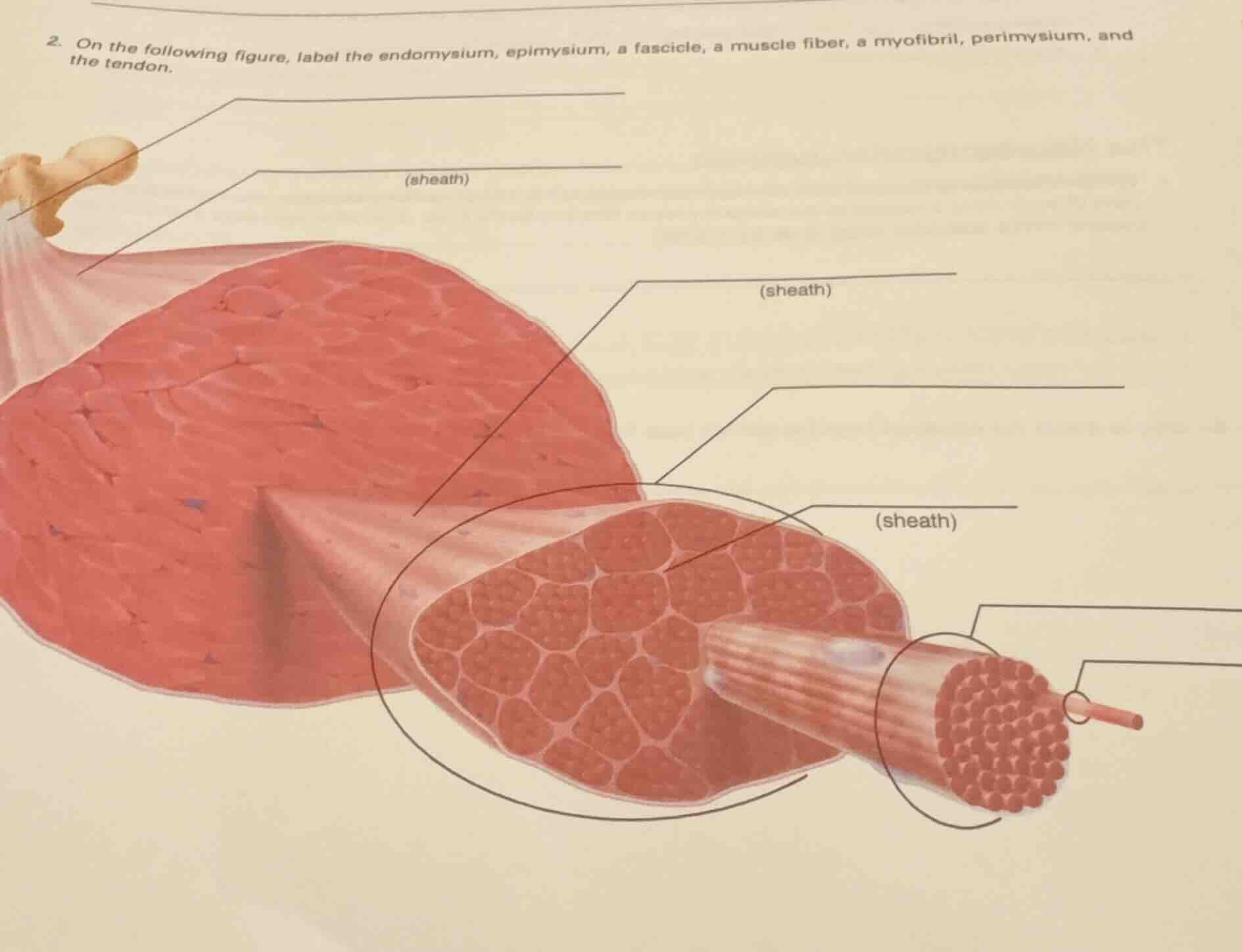
- on the following figure, label the endomysium, epimysium, a fascicle, a muscle fiber, a myofibril, perimysium, and the tendon.
(sheath)
(sheath)
(sheath)
Identify the structural hierarchy
We analyze the structural levels of a skeletal muscle from largest to smallest. The muscle connects to bone via a tendon. The outer muscle is wrapped in epimysium. Inside, fascicles are wrapped in perimysium. Each fascicle contains muscle fibers wrapped in endomysium. Each fiber contains myofibrils.
Map the pointer lines
We trace each pointer line from top-left to bottom-right:
- Top-left pointer: Points to the dense connective tissue connecting muscle to bone. This is the tendon.
- Second pointer from top-left: Points to the outer connective tissue sheath surrounding the entire muscle. This is the epimysium (sheath).
- Middle pointer pointing to the sheath of the pulled-out bundle: Points to the sheath surrounding a fascicle. This is the perimysium (sheath).
- Pointer pointing to the pulled-out bundle itself: This bundle is a fascicle.
- Pointer pointing to the sheath inside the fascicle surrounding an individual muscle cell: This is the endomysium (sheath).
- Pointer pointing to the single muscle cell pulled out from the fascicle: This is a muscle fiber.
- Bottom-right pointer pointing to the thread-like organelle pulled out from the fiber: This is a myofibril.
Relate to muscle characteristics
Using our knowledge of Skeletal Muscle Characteristics, we confirm that these nested layers of connective tissue sheaths provide structural support and channel blood vessels and nerves throughout the muscle.

Snap & solve any problem in the app
Get step-by-step solutions on Sovi AI
Photo-based solutions with guided steps
Explore more problems and detailed explanations
Based on the anatomical structure of a skeletal muscle from largest to smallest, the labels for the pointer lines (ordered generally from top-left to bottom-right) are:
- Tendon (top-most line, pointing to the connection to the bone)
- Epimysium (labeled as "sheath", pointing to the outer covering of the entire muscle)
- Perimysium (labeled as "sheath", pointing to the covering of the pulled-out fascicle)
- Fascicle (pointing to the pulled-out bundle of muscle fibers)
- Endomysium (labeled as "sheath", pointing to the covering of an individual muscle fiber within the fascicle)
- Muscle fiber (pointing to the single muscle cell pulled out from the bundle)
- Myofibril (pointing to the individual organelle strand pulled out from the muscle fiber)