QUESTION IMAGE
Question
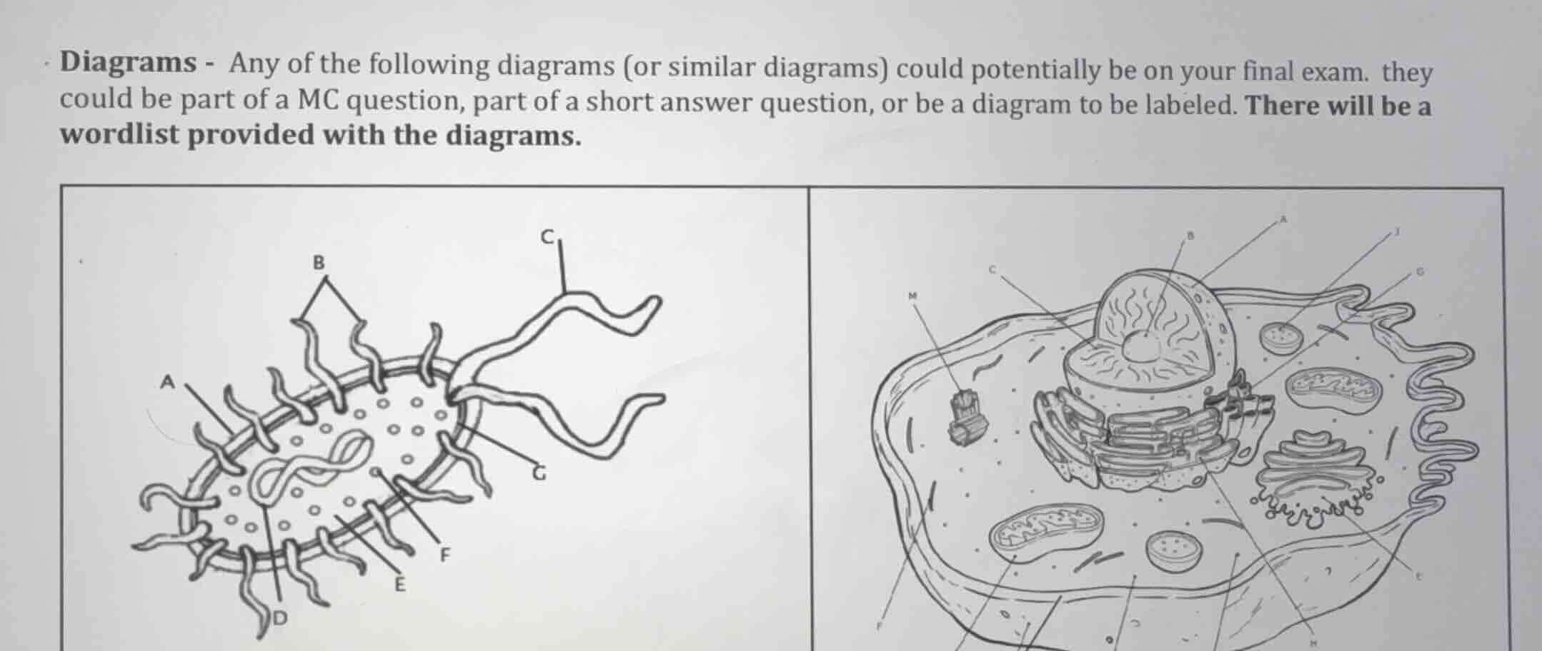
diagrams - any of the following diagrams (or similar diagrams) could potentially be on your final exam. they could be part of a mc question, part of a short answer question, or be a diagram to be labeled. there will be a wordlist provided with the diagrams.
Identify prokaryotic cell structures (left diagram)
- A: Capsule / Cell wall (outer protective layers)
- B: Pili / Fimbriae (hair-like appendages)
- C: Flagellum (tail-like structure for movement)
- D: Cell membrane / Plasma membrane (inner boundary layer)
- E: Ribosome (small dots in cytoplasm)
- F: Nucleoid / DNA (circular genetic material)
- G: Cytoplasm (fluid filling the cell)
Identify eukaryotic cell structures (right diagram)
- A: Nucleus (nuclear envelope/membrane)
- B: Nucleolus (dense region inside nucleus)
- C: Chromatin / DNA (inside nucleus)
- D: Rough Endoplasmic Reticulum (studded with ribosomes)
- E: Golgi apparatus (stacked membrane structure)
- F: Cytoplasm / Cytosol (fluid matrix)
- G: Lysosome / Peroxisome (small vesicle)
- H: Ribosome (free-floating or on ER)
- I: Mitochondrion (oval with folded inner membrane)
- J: Plasma membrane (outer boundary)
- K: Microtubules / Cytoskeleton (structural fibers)
- L: Centrosome / Centriole (barrel-shaped organelle)

Snap & solve any problem in the app
Get step-by-step solutions on Sovi AI
Photo-based solutions with guided steps
Explore more problems and detailed explanations
Question 1: Prokaryotic Cell Diagram (Left)
- A: Capsule (or Cell Wall)
- B: Pili (or Fimbriae)
- C: Flagellum
- D: Plasma Membrane
- E: Ribosome
- F: Nucleoid (DNA)
- G: Cytoplasm
Question 2: Eukaryotic Cell Diagram (Right)
- A: Nucleus (Nuclear Membrane)
- B: Nucleolus
- C: Chromatin (DNA)
- D: Rough Endoplasmic Reticulum
- E: Golgi Apparatus
- F: Cytoplasm (or Cytosol)
- G: Lysosome (or Peroxisome)
- H: Ribosome
- I: Mitochondrion
- J: Plasma Membrane
- K: Cytoskeleton (Microfilaments/Microtubules)
- L: Centriole (or Centrosome)