QUESTION IMAGE
Question
activity 17: quiz
module: nervous
topic: structures / type: combination
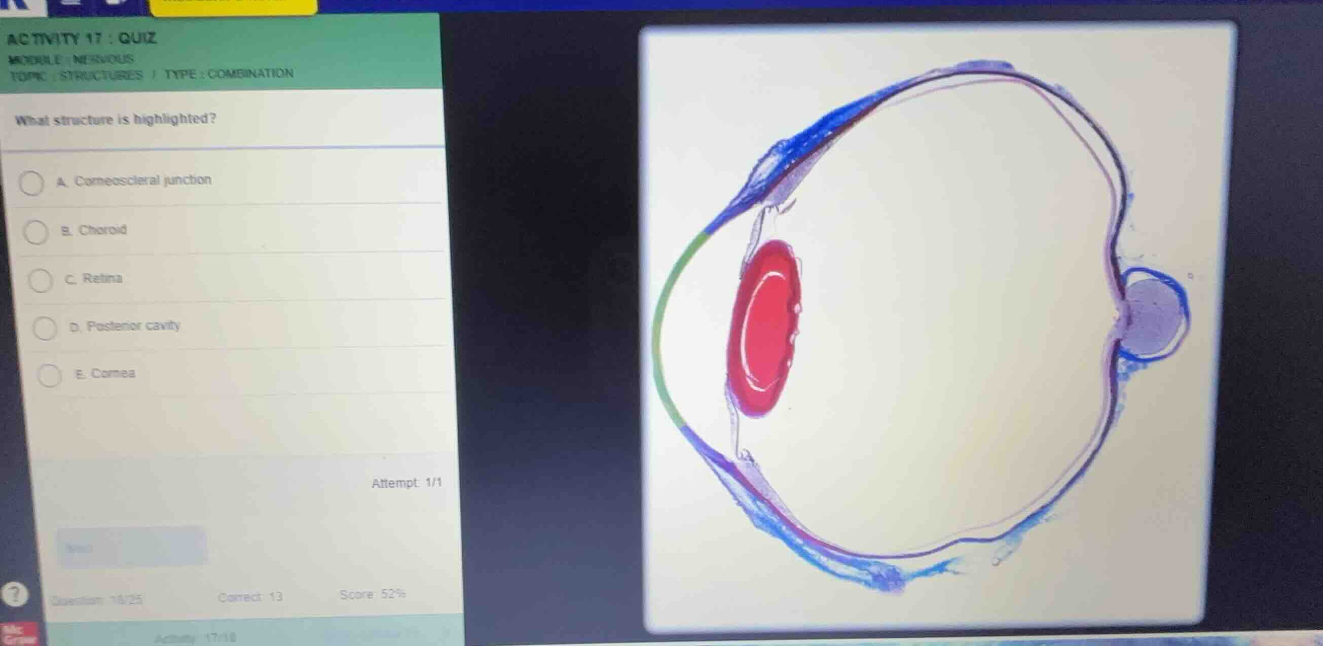
what structure is highlighted?
a. corneoscleral junction
b. choroid
c. retina
d. posterior cavity
e. cornea
Identify the highlighted structure
Using the Eye Anatomy and Retina Anatomy knowledge points
- The histological cross-section of the eye shows several color-coded structures:
- The green layer at the anterior represents the cornea.
- The red structure represents the lens.
- The blue outer layer represents the sclera.
- The thin pink/magenta layer lining the posterior inner surface of the eye represents the retina.
- The highlighted layer in pink/magenta is the innermost nervous tunic of the eye, which is the retina.
Match with options
Using the Ocular Histology knowledge point
- Option A: Corneoscleral junction (limbus) is located at the transition between the cornea and sclera.
- Option B: Choroid is the middle vascular layer (not highlighted here).
- Option C: Retina is the highlighted pink/magenta inner layer.
- Option D: Posterior cavity is the large space behind the lens.
- Option E: Cornea is the anterior transparent layer (highlighted in green).
- Therefore, the highlighted structure is the retina.

Snap & solve any problem in the app
Get step-by-step solutions on Sovi AI
Photo-based solutions with guided steps
Explore more problems and detailed explanations
- A. Corneoscleral junction
- B. Choroid
- C. Retina (Correct answer)
- D. Posterior cavity
- E. Cornea