QUESTION IMAGE
Question
a&p i
11 pts total
problem set 4.2: ch. 6, skeletal tissues
name(s)
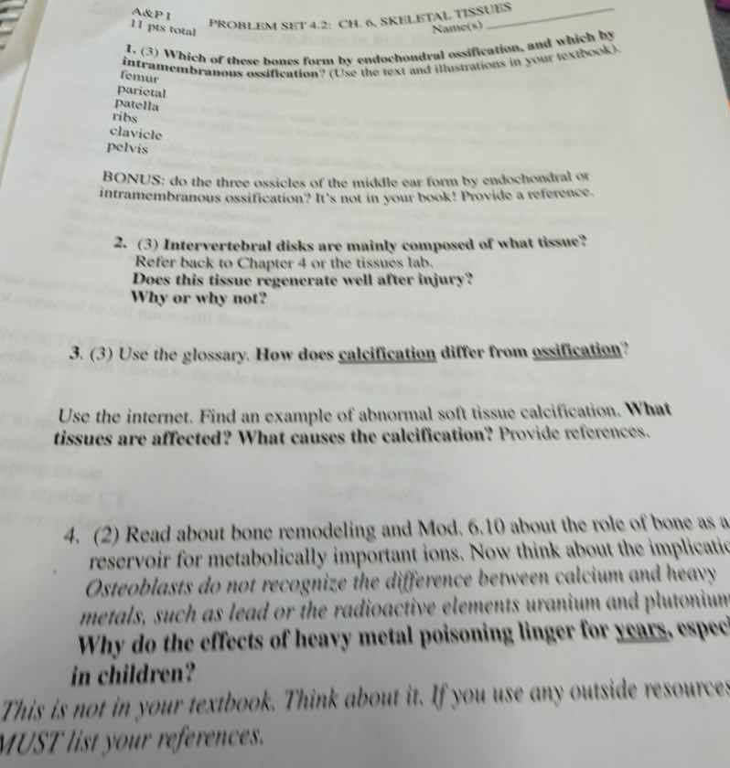
- (3) which of these bones form by endochondral ossification, and which by intramembranous ossification? (use the text and illustrations in your textbook).
femur
parietal
patella
ribs
clavicle
pelvis
bonus: do the three ossicles of the middle ear form by endochondral or intramembranous ossification? its not in your book! provide a reference.
- (3) intervertebral disks are mainly composed of what tissue?
refer back to chapter 4 or the tissues lab.
does this tissue regenerate well after injury?
why or why not?
- (3) use the glossary. how does calcification differ from ossification?
use the internet. find an example of abnormal soft tissue calcification. what tissues are affected? what causes the calcification? provide references.
- (2) read about bone remodeling and mod. 6.10 about the role of bone as a reservoir for metabolically important ions. now think about the implications. osteoblasts do not recognize the difference between calcium and heavy metals, such as lead or the radioactive elements uranium and plutonium. why do the effects of heavy metal poisoning linger for years, especially in children?
this is not in your textbook. think about it. if you use any outside resources, must list your references.
🆕 New Concept Discovered: Bone Development and Remodeling
How bones form, repair, and store minerals
---
Step 1: Bone Ossification Types (Question 1 & Bonus)
Bone formation (ossification) occurs via two main pathways:
- Intramembranous ossification: Bone develops directly from sheets of mesenchymal (undifferentiated) connective tissue. This process primarily forms flat bones of the skull, the mandible, and most of the clavicles.
- Endochondral ossification: Bone develops by replacing a pre-existing hyaline cartilage model. This process forms most of the bones in the skeleton, including long bones, ribs, and the pelvis.
Classification of the listed bones:
- Femur: Endochondral ossification (it is a long bone of the leg).
- Parietal: Intramembranous ossification (it is a flat bone of the skull).
- Patella: Endochondral ossification (it develops as a sesamoid bone within a cartilage model/tendon).
- Ribs: Endochondral ossification (they develop from cartilaginous precursors).
- Clavicle: Intramembranous ossification (specifically, the medial and lateral ends form primarily via intramembranous pathways, though it is unique in having some endochondral characteristics at its medial end; standard introductory A&P textbooks classify it under intramembranous).
- Pelvis: Endochondral ossification (it forms from cartilage models of the ilium, ischium, and pubis).
BONUS: Middle Ear Ossicles (Malleus, Incus, Stapes)
The three auditory ossicles form via endochondral ossification. They develop from the cartilages of the first and second pharyngeal arches (Meckel's cartilage for the malleus and incus, and Reichert's cartilage for the stapes).
- Reference: Anthwal, N., & Thompson, H. (2016). "The development of the mammalian outer and middle ear." Journal of Anatomy, 228(2), 217-230.
---
Step 2: Intervertebral Disks Composition and Regeneration (Question 2)
Tissue Composition
Intervertebral disks are mainly composed of fibrocartilage (specifically making up the outer ring, the annulus fibrosus, which surrounds the gelatinous inner core, the nucleus pulposus).
Regeneration Ability
No, fibrocartilage does not regenerate well after injury.
Why or Why Not?
Cartilage, including fibrocartilage, is avascular (lacks a direct blood supply). Because there are no blood vessels running through the tissue, it is extremely difficult for essential nutrients, oxygen, and reparative cells (like fibroblasts or chondrocytes) to reach the injured site. Healing relies on slow diffusion from surrounding tissues, making the regenerative process incredibly slow and often incomplete, typically resulting in permanent scarring or degeneration rather than functional tissue repair.
---
Step 3: Calcification vs. Ossification & Abnormal Soft Tissue Calcification (Question 3)
Calcification vs. Ossification
- Calcification: The deposition and buildup of calcium salts in any tissue (either bony or soft tissue). It is a chemical process of hardening.
- Ossification: The biological process of actual bone formation, which requires the active work of specialized bone cells (osteoblasts laying down an organic osteoid matrix that is subsequently mineralized). Calcification is simply one step within the broader process of ossification.
Abnormal Soft Tissue Calcification Example
- Example: Calcific Tendonitis (specifically of the rotator cuff in the shoulder).
- Tissues Affected: Tendons (dense regular connective tissue), most commonly th…

Snap & solve any problem in the app
Get step-by-step solutions on Sovi AI
Photo-based solutions with guided steps
Explore more problems and detailed explanations
1. Ossification Classification
- Endochondral ossification: Femur, Patella, Ribs, Pelvis
- Intramembranous ossification: Parietal, Clavicle
- BONUS: The three middle ear ossicles form by endochondral ossification. (Reference: Anthwal & Thompson, 2016, J. Anat.)
2. Intervertebral Disks
- Tissue: Fibrocartilage.
- Regeneration: No, it does not regenerate well.
- Reason: It is avascular (lacks blood vessels), which severely limits the delivery of nutrients, oxygen, and progenitor cells necessary for repair.
3. Calcification vs. Ossification
- Difference: Calcification is the physical deposition of calcium salts in any tissue, whereas ossification is the biological process of bone tissue formation by osteoblasts.
- Abnormal Calcification Example: Calcific Tendonitis of the rotator cuff.
- Tissues Affected: Tendons (dense regular connective tissue).
- Causes: Localized hypoxia, microtrauma, or cellular misdifferentiation leading to calcium crystal deposition. (References: Uhthoff & Loehr, 1997; Oliva et al., 2016)
4. Heavy Metal Poisoning in Bones
- Why it lingers: Osteoblasts mistake heavy metals (like lead, uranium, and plutonium) for calcium due to chemical similarity and lock them into the bone's mineral matrix. These toxins are only slowly released back into the body over years during normal bone remodeling.
- Why children are affected more: Children have rapid bone growth rates, meaning they absorb these metals into their skeletons much faster. The continuous release of these stored toxins during development causes long-term damage to their highly sensitive, developing nervous systems.