QUESTION IMAGE
Question
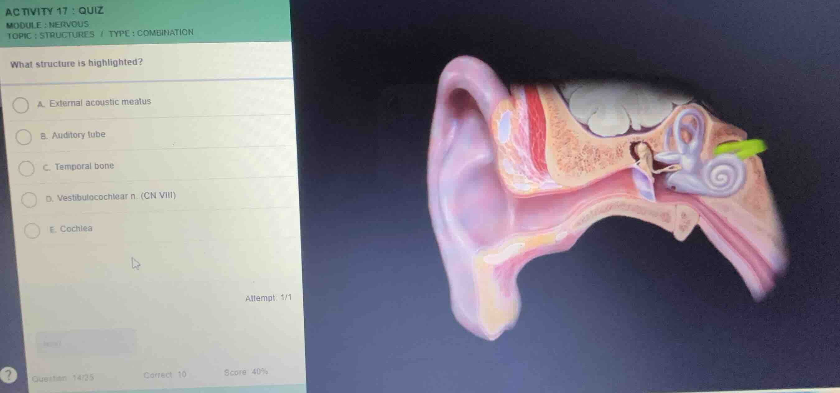
what structure is highlighted?
a. external acoustic meatus
b. auditory tube
c. temporal bone
d. vestibulocochlear n. (cn viii)
e. cochlea
Identify the highlighted structure
Using the Auditory System Anatomy knowledge point
- The anatomical diagram shows a cross-section of the human ear, including the outer, middle, and inner ear structures.
- On the right side of the diagram, emerging from the inner ear (specifically from the vestibule and cochlea), there is a prominent nerve highlighted in bright green.
- This nerve is the vestibulocochlear nerve (Cranial Nerve VIII), which carries sensory information for both hearing and balance to the brain.
Evaluate the options
- A. External acoustic meatus: This is the ear canal leading from the auricle to the tympanic membrane, which is not highlighted.
- B. Auditory tube: Also known as the Eustachian tube, this connects the middle ear to the nasopharynx and is located inferiorly, not highlighted.
- C. Temporal bone: This is the surrounding bone structure of the middle and inner ear, shown in beige/brown, not highlighted.
- D. Vestibulocochlear n. (CN VIII): This matches the bright green highlighted nerve bundle exiting the inner ear.
- E. Cochlea: This is the snail-shell-like structure of the inner ear, shown in grey/white, but is not the highlighted green structure itself.

Snap & solve any problem in the app
Get step-by-step solutions on Sovi AI
Photo-based solutions with guided steps
Explore more problems and detailed explanations
- (A) External acoustic meatus
- (B) Auditory tube
- (C) Temporal bone
- (D) Vestibulocochlear n. (CN VIII) (Correct answer)
- (E) Cochlea