QUESTION IMAGE
Question
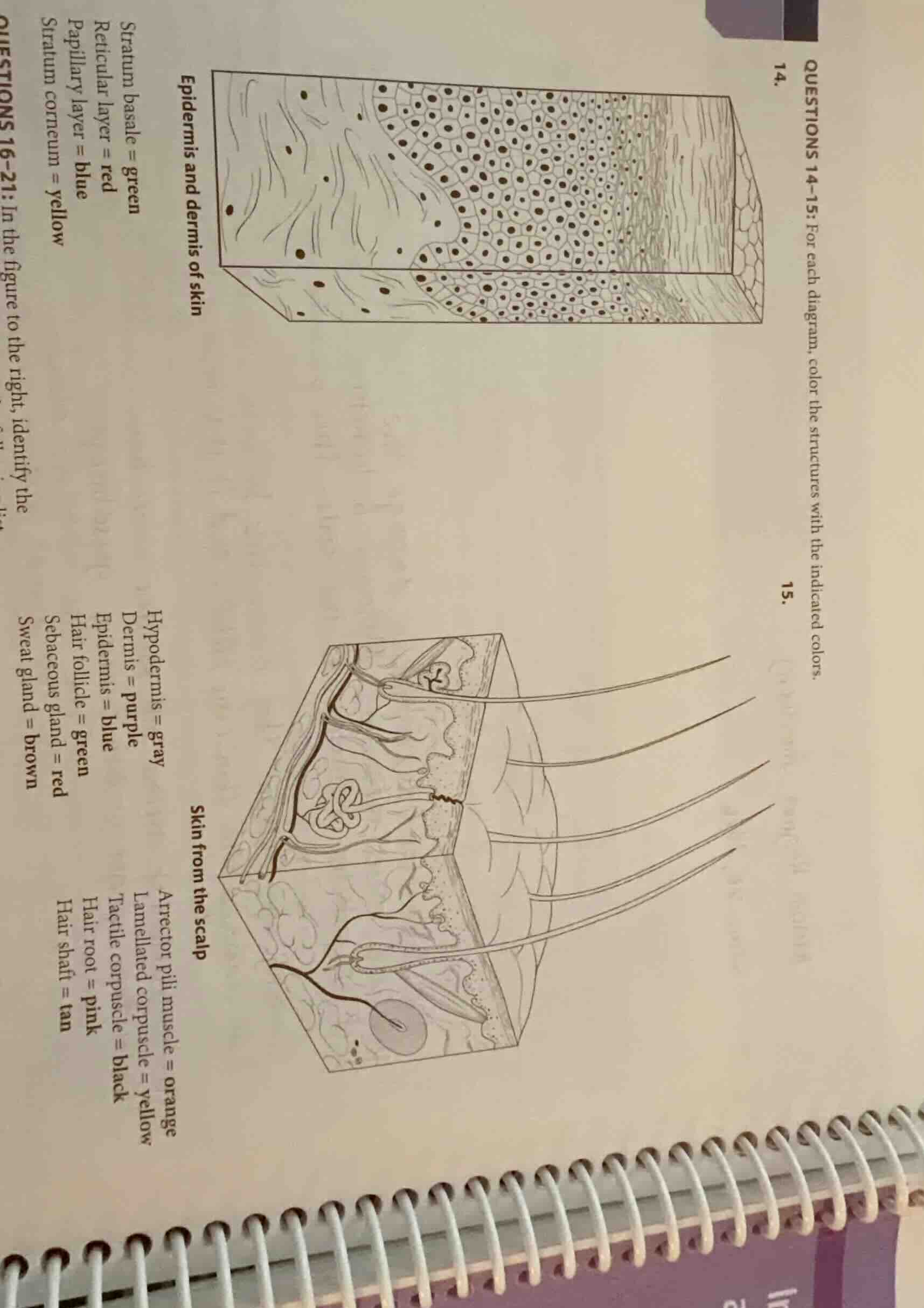
questions 14-15: for each diagram, color the structures with the indicated colors.
- epidermis and dermis of skin
stratum basale = green
reticular layer = red
papillary layer = blue
stratum corneum = yellow
- skin from the scalp
hypodermis = gray
dermis = purple
epidermis = blue
hair follicle = green
sebaceous gland = red
sweat gland = brown
arrector pili muscle = orange
lamellated corpuscle = yellow
tactile corpuscle = black
hair root = pink
hair shaft = tan
🆕 New Concept Discovered: Integumentary System Anatomy
Understanding the layers and structures of the skin.
Step 1: Identify structures in Diagram 14 (Epidermis and dermis of skin)
To color the diagram correctly, we must locate each layer of the epidermis and dermis:
- Stratum basale (Green): This is the deepest, single-cell layer of the epidermis, resting directly on the wavy basement membrane that separates the epidermis from the dermis.
- Stratum corneum (Yellow): This is the outermost, thick layer of flat, dead, keratinized cells at the very top surface of the skin.
- Papillary layer of dermis (Blue): This is the upper region of the dermis directly beneath the epidermis, characterized by finger-like projections (dermal papillae) that indent the epidermis.
- Reticular layer of dermis (Red): This is the deeper, thicker layer of the dermis beneath the papillary layer, containing dense irregular connective tissue fibers.
Step 2: Identify structures in Diagram 15 (Skin from the scalp)
To color the scalp diagram correctly, we locate the main layers and specialized accessory structures:
- Epidermis (Blue): The thin, outermost cellular layer of the skin.
- Dermis (Purple): The thick middle layer of connective tissue beneath the epidermis containing glands, hair follicles, and sensory receptors.
- Hypodermis (Gray): The deepest layer (subcutaneous layer) containing adipose (fat) tissue, located at the very bottom of the block.
- Hair follicle (Green): The sheath-like structure made of epithelial and connective tissue that surrounds the hair root.
- Hair root (Pink): The portion of the hair anchored deep within the hair follicle, below the skin surface.
- Hair shaft (Tan): The portion of the hair that projects above the skin surface.
- Sebaceous gland (Red): The multi-lobed, oil-producing glands connected directly to the upper part of the hair follicles.
- Sweat gland (Brown): The coiled, tubular gland located deep in the dermis with a duct leading to a pore on the skin surface.
- Arrector pili muscle (Orange): The tiny band of smooth muscle connecting the hair follicle to the upper dermis.
- Tactile corpuscle (Black): Small, oval sensory receptors located high up in the dermal papillae (just beneath the epidermis) for light touch.
- Lamellated corpuscle (Yellow): Large, onion-like layered sensory receptors located deep in the dermis or hypodermis for deep pressure.

Snap & solve any problem in the app
Get step-by-step solutions on Sovi AI
Photo-based solutions with guided steps
Explore more problems and detailed explanations
Follow this guide to color the diagrams:
Diagram 14: Epidermis and dermis of skin
- Stratum basale: Color the single row of cells at the very bottom boundary of the epidermis green.
- Stratum corneum: Color the thick, outermost layer of flat cells at the top surface yellow.
- Papillary layer: Color the wavy upper region of the dermis containing the dermal papillae blue.
- Reticular layer: Color the deeper, fibrous region of the dermis below the papillae red.
---
Diagram 15: Skin from the scalp
- Epidermis: Color the entire thin outer layer of the skin blue.
- Dermis: Color the thick middle layer containing the glands and follicles purple.
- Hypodermis: Color the bottom-most layer containing the rounded fat cells gray.
- Hair follicle: Color the sheath surrounding the hair root green.
- Hair root: Color the lower portion of the hair embedded inside the follicle pink.
- Hair shaft: Color the long strands of hair extending out above the skin surface tan.
- Sebaceous gland: Color the multi-lobed glands attached to the side of the hair follicles red.
- Sweat gland: Color the coiled, tube-like gland structure and its duct brown.
- Arrector pili muscle: Color the small diagonal muscle band attached to the hair follicle orange.
- Tactile corpuscle: Color the small, oval touch receptors in the dermal papillae black.
- Lamellated corpuscle: Color the large, layered pressure receptor deep in the dermis/hypodermis yellow.