QUESTION IMAGE
Question
Question was provided via image upload.
Identify the target anatomical structure
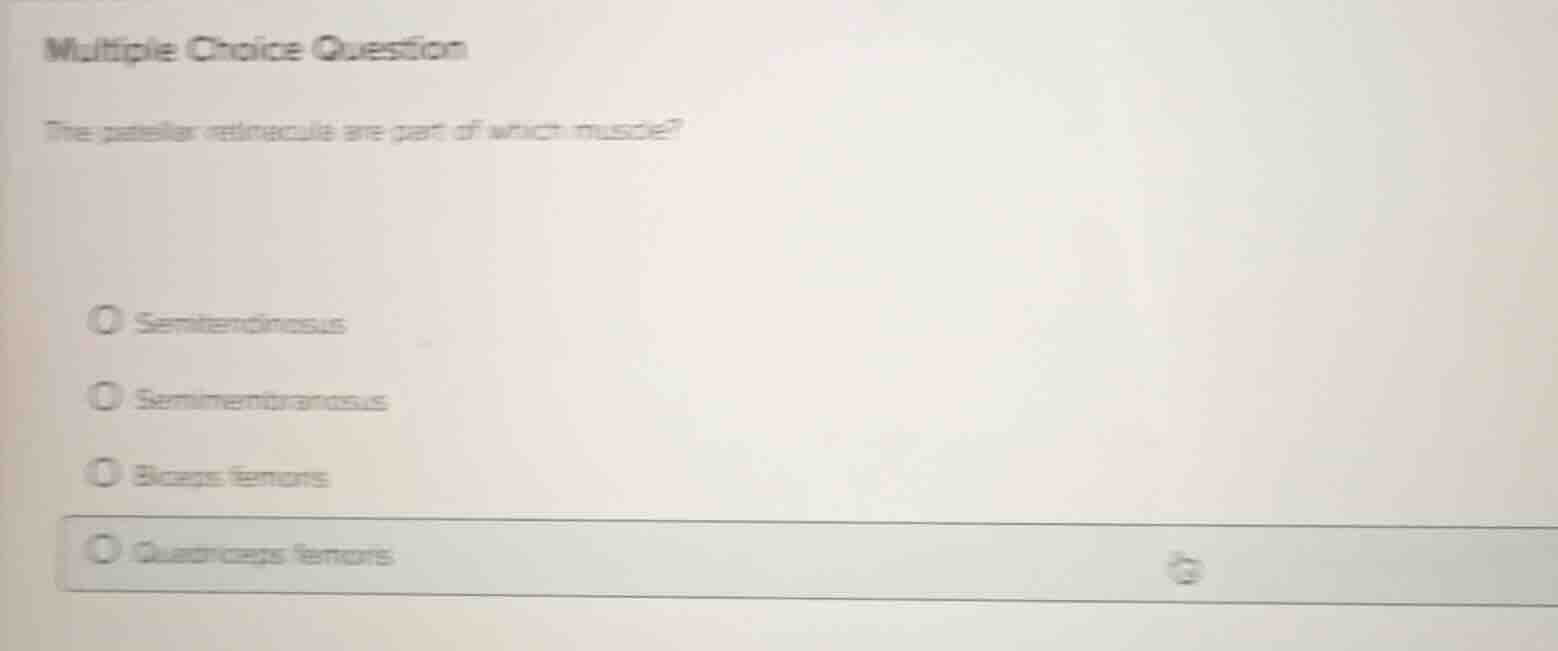
The question asks about the patellar retinacula (medial and lateral patellar retinacula) and which muscle tendon they originate from or are extensions of.
Analyze the relationship to knee joint anatomy
Using the Knee Joint Anatomy knowledge point, we recall that the patellar retinacula are fibrous expansions that support the knee joint anteriorly and help keep the patella aligned.
Trace the muscular connections
The medial and lateral patellar retinacula are formed by aponeurotic expansions of the vastus medialis and vastus lateralis muscles, which are key components of the quadriceps femoris muscle group.
Evaluate the given options
- Semitendinosus: A hamstring muscle on the posterior thigh.
- Semimembranosus: Another posterior hamstring muscle.
- Biceps femoris: The lateral posterior hamstring muscle.
- Quadriceps femoris: The anterior thigh muscle group whose tendon inserts into the patella and continues as the patellar ligament, with lateral expansions forming the retinacula.
Formulate the final selection
The patellar retinacula are structural continuations of the quadriceps femoris tendon. Therefore, the correct option is Quadriceps femoris.

Snap & solve any problem in the app
Get step-by-step solutions on Sovi AI
Photo-based solutions with guided steps
Explore more problems and detailed explanations
- Semitendinosus
- Semimembranosus
- Biceps femoris
- Quadriceps femoris (Correct answer)