QUESTION IMAGE
Question
Question was provided via image upload.
🆕 New Concept Discovered: Connective Tissue Histology
Identifying specialized supportive tissues under the microscope.
Step 1: Identify the Left Image
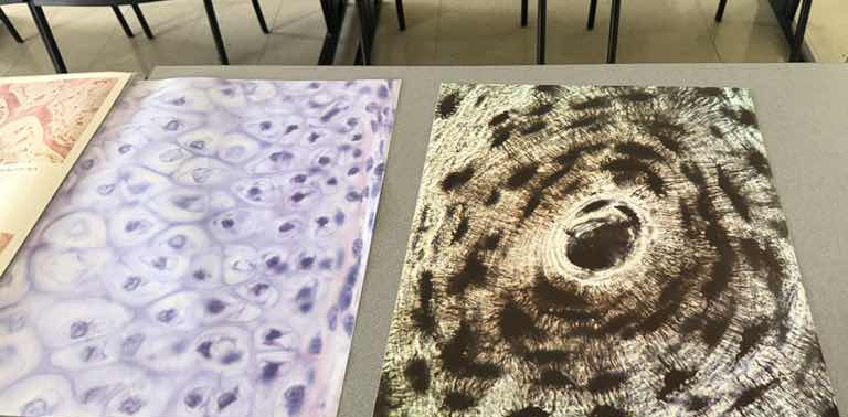
The image on the left shows cells residing in small spaces called lacunae, surrounded by a smooth, glassy extracellular matrix.
- Key Features: Chondrocytes (cartilage cells) located within lacunae, often appearing in pairs or small groups (isogenous groups). The matrix is abundant and lacks visible fibers under standard light microscopy.
- Tissue Type: Hyaline Cartilage
Step 2: Identify the Right Image
The image on the right shows a highly organized, circular structure resembling tree rings.
- Key Features: A large central opening (Haversian canal) surrounded by concentric rings of matrix (lamellae). Small, dark, spider-like spaces (lacunae containing osteocytes) are arranged along the rings, with tiny radiating channels (canaliculi) connecting them.
- Tissue Type: Compact Bone (specifically, an osteon or Haversian system)

Snap & solve any problem in the app
Get step-by-step solutions on Sovi AI
Photo-based solutions with guided steps
Explore more problems and detailed explanations
- Left Image: Hyaline Cartilage
- Right Image: Compact Bone (Osteon)