QUESTION IMAGE
Question
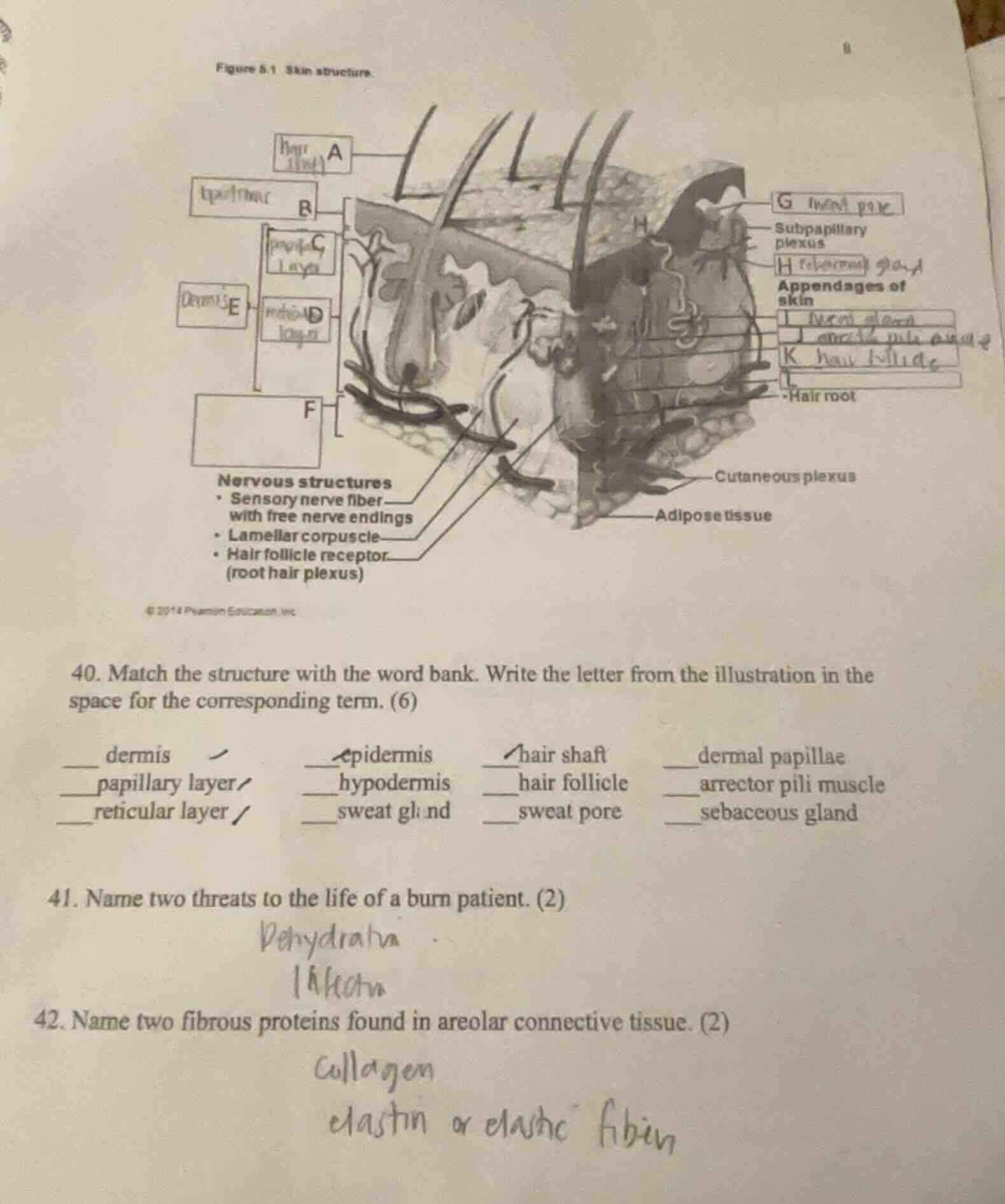
figure 5.1 skin structure.
nervous structures
- sensory nerve fiber with free nerve endings
- lamellar corpuscle
- hair follicle receptor (root hair plexus)
- subpapillary plexus
appendages of skin
- hair root
- cutaneous plexus
- adipose tissue
- match the structure with the word bank. write the letter from the illustration in the space for the corresponding term. (6)
___ dermis
___ epidermis
___ hair shaft
___ dermal papillae
___ papillary layer
___ hypodermis
___ hair follicle
___ arrector pili muscle
___ reticular layer
___ sweat gland
___ sweat pore
___ sebaceous gland
- name two threats to the life of a burn patient. (2)
- name two fibrous proteins found in areolar connective tissue. (2)
Identify skin structures from the diagram labels
- A: Hair shaft (the part of the hair projecting above the skin surface)
- B: Epidermis (the outermost layer of the skin)
- C: Papillary layer (the superficial layer of the dermis)
- D: Reticular layer (the deeper layer of the dermis)
- E: Dermis (the layer beneath the epidermis containing C and D)
- F: Hypodermis (the subcutaneous layer beneath the dermis)
- G: Sweat pore (the opening of a sweat gland duct on the skin surface)
- H: Sebaceous gland (oil gland connected to the hair follicle)
- I: Arrector pili muscle (smooth muscle attached to the hair follicle)
- J: Sweat gland (sudoriferous gland in the dermis)
- K: Hair follicle (sheath surrounding the hair root)
Identify threats to a burn patient
- Dehydration: Loss of the protective skin barrier leads to massive fluid loss, causing hypovolemic shock.
- Infection: The loss of the physical barrier allows pathogens to enter, leading to sepsis.
Identify fibrous proteins in areolar connective tissue
- Collagen fibers: Provide tensile strength.
- Elastic fibers (or elastin): Provide elasticity and resilience.
- Reticular fibers: Form a supportive meshwork.

Snap & solve any problem in the app
Get step-by-step solutions on Sovi AI
Photo-based solutions with guided steps
Explore more problems and detailed explanations
Question 40
- E dermis
- B epidermis
- A hair shaft
- C dermal papillae (or papillary layer)
- C papillary layer
- F hypodermis
- K hair follicle
- I arrector pili muscle
- D reticular layer
- J sweat gland
- G sweat pore
- H sebaceous gland
Question 41
- Dehydration (extreme fluid loss leading to hypovolemic shock)
- Infection (loss of skin barrier leading to bacterial invasion and sepsis)
Question 42
- Collagen (collagen fibers)
- Elastin (elastic fibers)