QUESTION IMAGE
Question
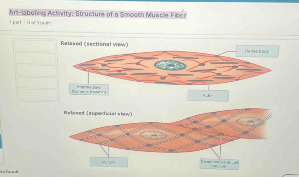
art-labeling activity: structure of a smooth muscle fiber
1 part | 0 of 1 point
relaxed (sectional view)
dense body
intermediate filaments (desmin)
actin
relaxed (superficial view)
myosin
dense bodies at cell junction
🆕 New Concept Discovered: Smooth Muscle Microanatomy
Understanding how non-striated contractile units organize.
Step 1: Identify the missing labels
The image shows an "Art-labeling Activity: Structure of a Smooth Muscle Fiber" with five empty draggable boxes on the left. However, looking closely at the diagram, all the pointer lines pointing to the structures are already labeled:
- Intermediate filaments (desmin)
- Dense body
- Actin
- Myosin
- Dense bodies at cell junction
In standard anatomy homework platforms (such as MasteringA&P), the list of labels on the left contains the terms that need to be dragged into the empty slots on the diagram. Since the diagram in this screenshot is already fully labeled, the five boxes on the left represent the pool of labels you drag from.
Step 2: Match the labels to their corresponding structures
If you need to drag the labels from the left column into their correct positions on an unlabeled version of this diagram, here is the correct mapping:
- Intermediate filaments (desmin): Points to the thin structural network lines running longitudinally through the relaxed sectional view.
- Dense body: Points to the dark, circular anchor points distributed throughout the sarcoplasm in the sectional view.
- Actin: Points to the thin contractile filaments (red lines) in the sectional view.
- Myosin: Points to the thick contractile filaments (represented by the lattice lines on the superficial view).
- Dense bodies at cell junction: Points to the dark spots where adjacent smooth muscle cells meet and anchor to one another.

Snap & solve any problem in the app
Get step-by-step solutions on Sovi AI
Photo-based solutions with guided steps
Explore more problems and detailed explanations
The labels shown in the left column correspond to the following structures on the diagram:
- Intermediate filaments (desmin) (points to the internal structural network)
- Dense body (points to the dark internal anchoring spots)
- Actin (points to the thin contractile filaments)
- Myosin (points to the thick contractile filaments on the surface lattice)
- Dense bodies at cell junction (points to the connection points between adjacent cells)