QUESTION IMAGE
Question
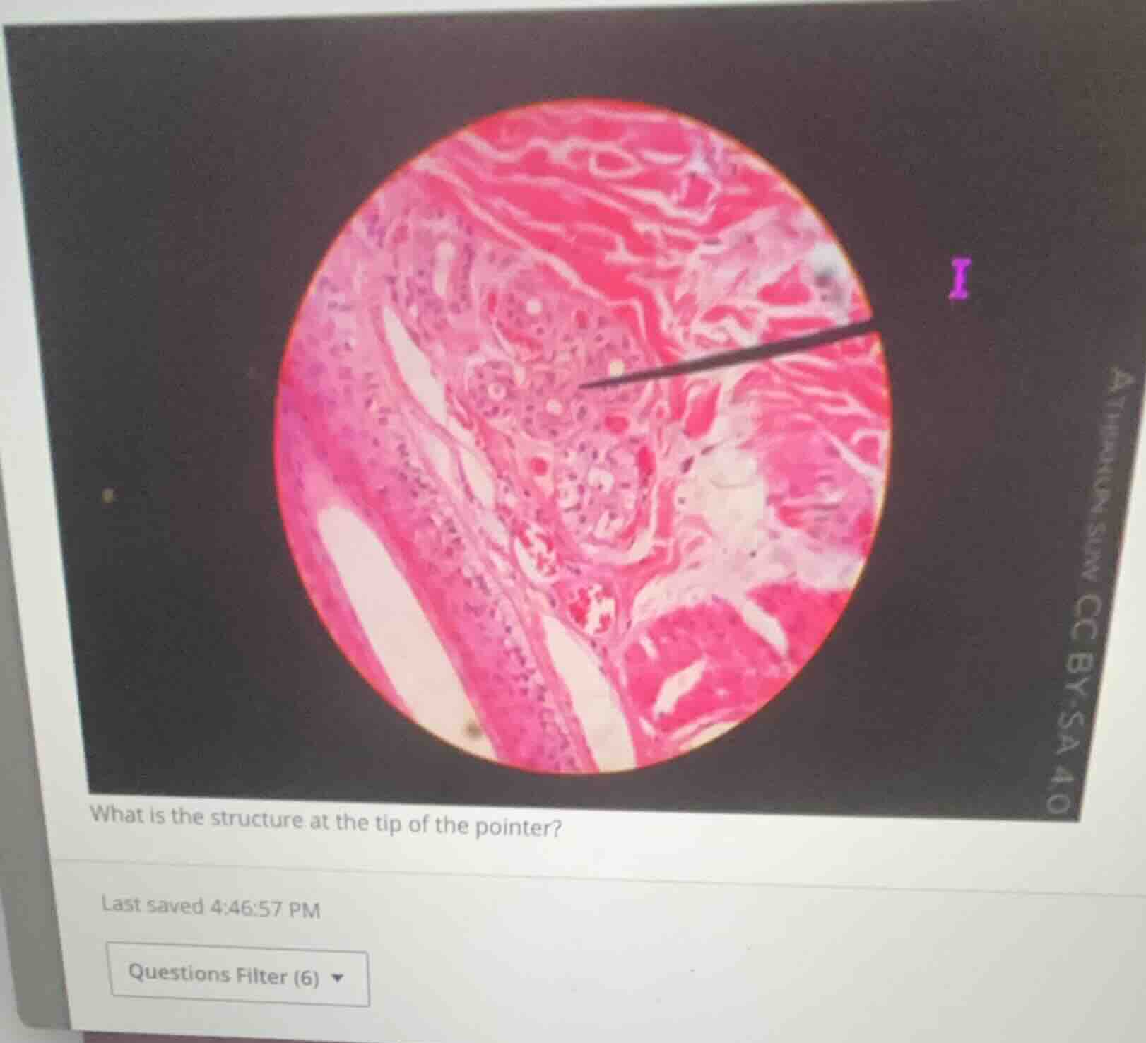
what is the structure at the tip of the pointer?
🆕 New Concept Discovered: Histological Identification of Skin Appendages
Recognizing glands and hair follicles in integumentary tissue.
Step 1: Analyze the tissue context
The image shows a high-magnification histological section of skin (integumentary system) stained with Hematoxylin and Eosin (H&E).
- On the left, we can see a hair follicle cut longitudinally/obliquely.
- Surrounding the structures is dense irregular connective tissue (dermis), stained bright pink due to collagen fibers.
Step 2: Identify the pointed structure
The pointer labeled 1 (or "I") points directly to a cluster of coiled, tubular epithelial structures located in the dermis near the hair follicle.
- These structures consist of simple cuboidal epithelial cells surrounding small, clear central lumens.
- This coiled, tubular appearance is highly characteristic of a sweat gland (specifically, a merocrine/eccrine sweat gland) cut in various cross-sections.

Snap & solve any problem in the app
Get step-by-step solutions on Sovi AI
Photo-based solutions with guided steps
Explore more problems and detailed explanations
Sweat gland (specifically, an eccrine/merocrine sweat gland)