QUESTION IMAGE
Question
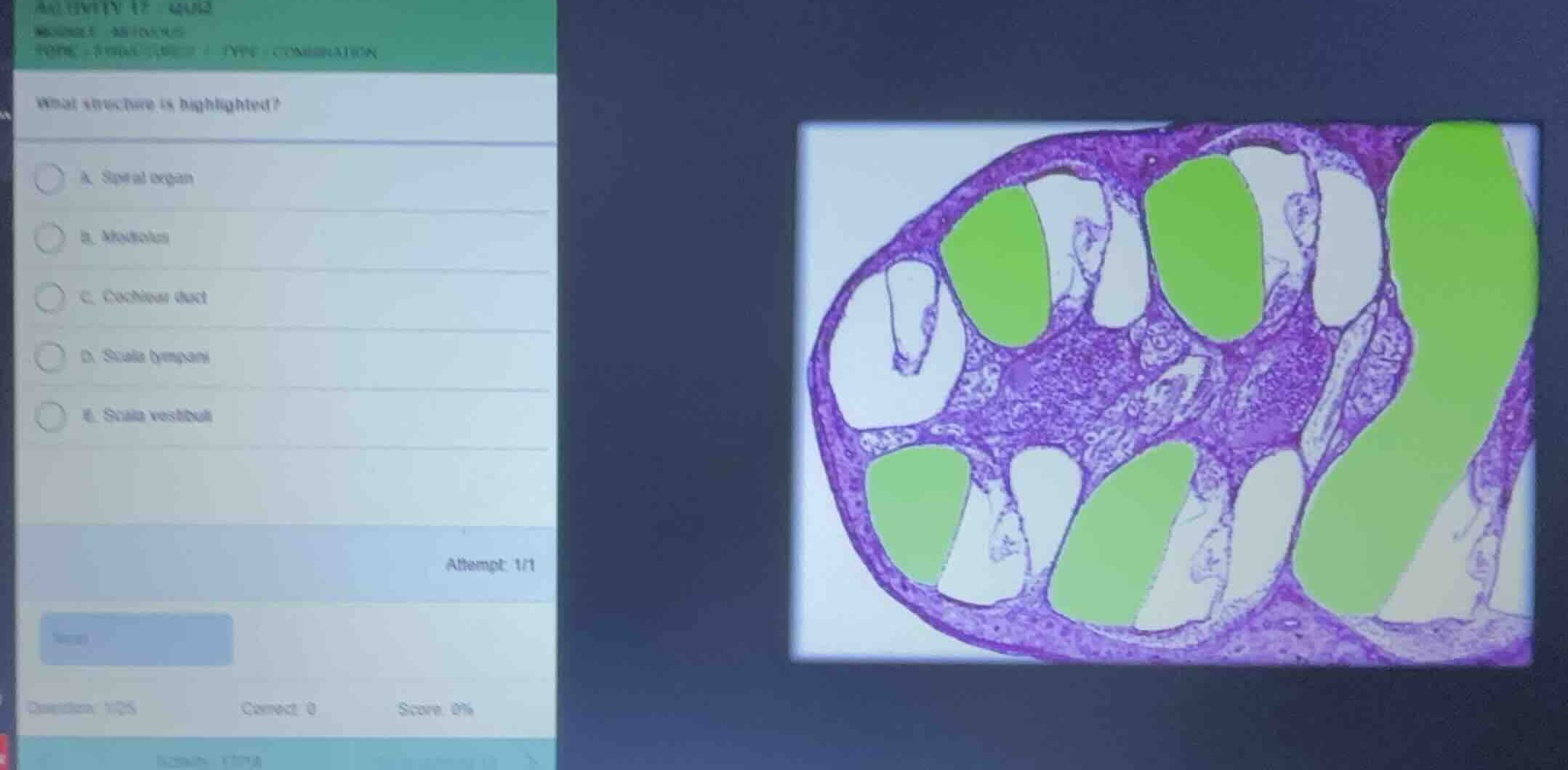
what structure is highlighted?
a. spiral organ
b. modiolus
c. cochlear duct
d. scala tympani
e. scala vestibuli
Identify the histological section
The image shows a cross-section of the cochlea of the inner ear. The cochlea is a spiral, bony canal divided into three fluid-filled chambers (scalae) that wrap around a central bony core called the modiolus.
Analyze the highlighted regions
The green highlighted regions correspond to the uppermost chamber in each turn of the cochlear canal.
Differentiate the cochlear chambers
Using our knowledge of Auditory System Anatomy, we can identify the three main compartments within each turn of the cochlea:
- Scala vestibuli: The superior chamber, which is filled with perilymph. In this section, it is located on the upper side of each turn, separated from the middle chamber by the thin vestibular (Reissner's) membrane. This matches the green highlighted areas.
- Cochlear duct (Scala media): The middle, triangular chamber filled with endolymph, containing the spiral organ (organ of Corti).
- Scala tympani: The inferior chamber, filled with perilymph, separated from the cochlear duct by the basilar membrane.
Since the highlighted green regions represent the superior chambers of the cochlear turns, the highlighted structure is the scala vestibuli.

Snap & solve any problem in the app
Get step-by-step solutions on Sovi AI
Photo-based solutions with guided steps
Explore more problems and detailed explanations
- A. Spiral organ
- B. Modiolus
- C. Cochlear duct
- D. Scala tympani
- E. Scala vestibuli (Correct answer)