QUESTION IMAGE
Question
what structure is highlighted?
a. optic tract
b. posterior cavity
c. posterior chamber
d. sclera
e. optic n. (cn ii)
Identify highlighted structure
Using the Eye Anatomy and Neural Components knowledge points
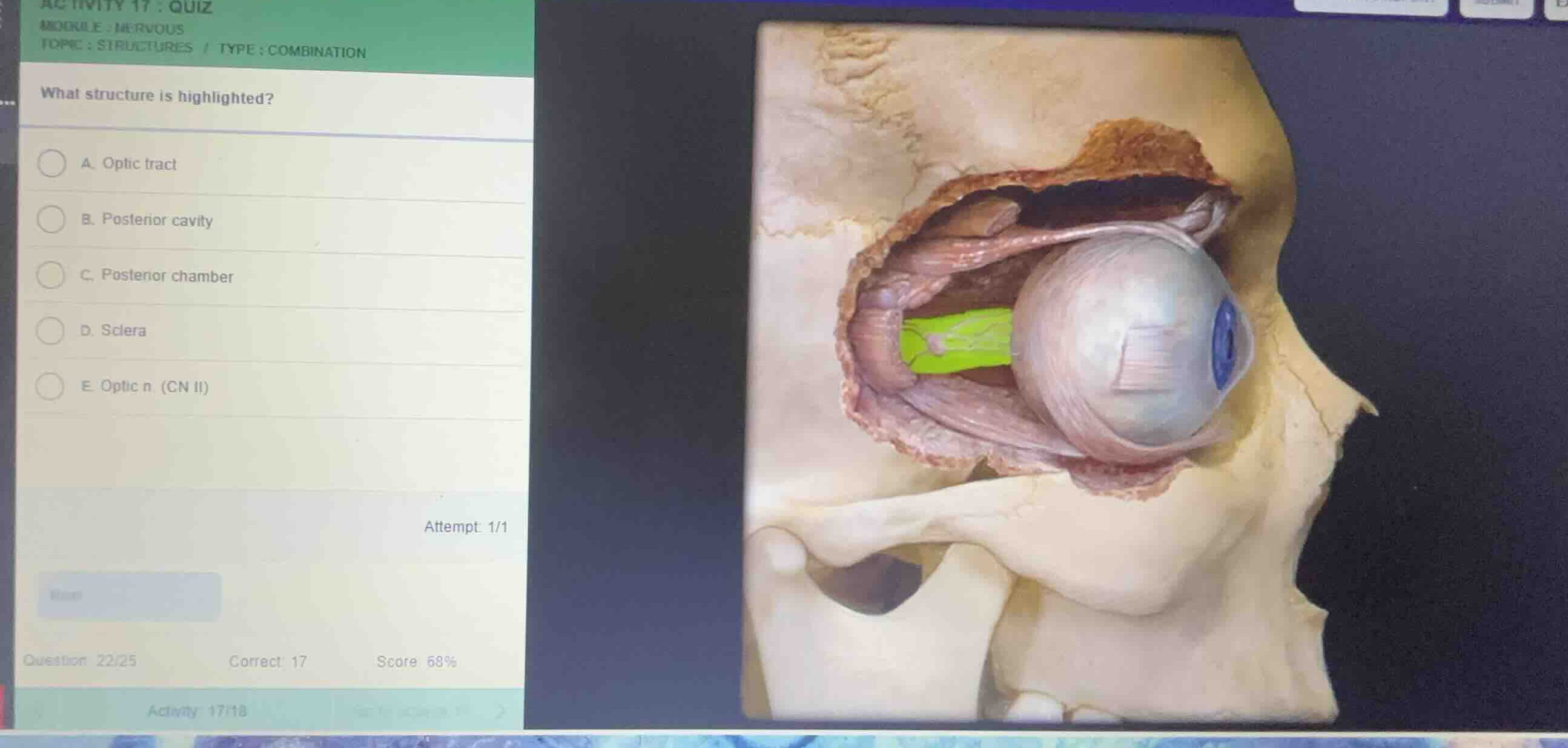
- The 3D anatomical model shows a lateral/sagittal view of the right orbit.
- The eyeball (bulbus oculi) is situated anteriorly.
- Extending posteriorly from the back of the eyeball through the optic canal is a prominent, thick nerve highlighted in bright green.
- This structure is the optic nerve, also known as Cranial Nerve II (CN II).
Evaluate the options
- A. Optic tract: The optic tract is located posterior to the optic chiasm inside the cranial cavity, not within the orbit.
- B. Posterior cavity: This refers to the vitreous chamber inside the eyeball itself.
- C. Posterior chamber: This is a small space inside the eye between the iris and the lens.
- D. Sclera: This is the white outer layer of the eyeball, not the nerve extending behind it.
- E. Optic n. (CN II): This correctly identifies the highlighted cranial nerve exiting the posterior aspect of the eye.

Snap & solve any problem in the app
Get step-by-step solutions on Sovi AI
Photo-based solutions with guided steps
Explore more problems and detailed explanations
- (A) Optic tract
- (B) Posterior cavity
- (C) Posterior chamber
- (D) Sclera
- (E) Optic n. (CN II) (Correct answer)