QUESTION IMAGE
Question
what structure is highlighted?
a. basilar membrane
b. nerve fibers of cochlea
c. stria vascularis
d. scala vestibuli
e. scala tympani
Identify the histological section
Using the Cochlea Histology and Cochlea Histology Identification knowledge points
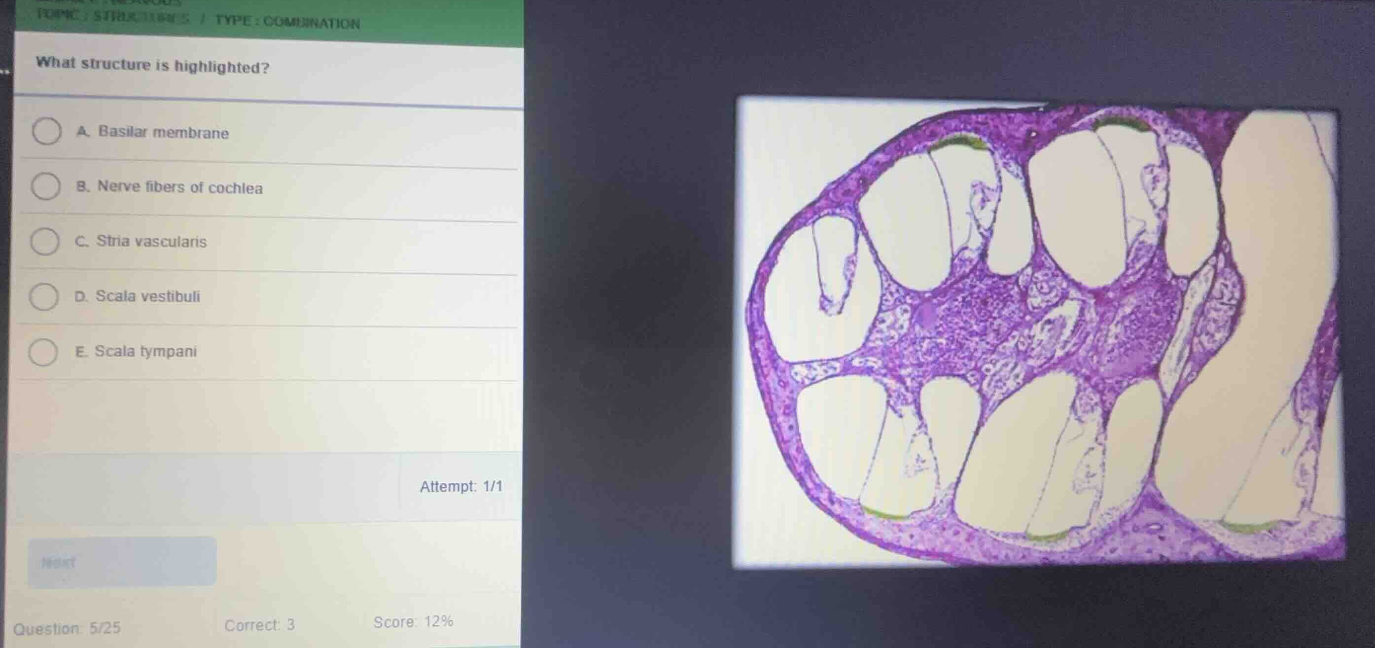
The image shows a cross-section of the bony cochlea, displaying multiple turns of the cochlear duct (scala media), scala vestibuli, and scala tympani.
Analyze the highlighted regions
Using the Cochlea Histology and Cochlea Histology Identification knowledge points
The highlighted regions (colored in green) are located on the outer wall of the cochlear duct (scala media). This specialized, highly vascularized pseudostratified epithelium is responsible for producing endolymph.
Match with the correct anatomical structure
Using the Cochlea Histology and Cochlea Histology Identification knowledge points
The vascularized tissue lining the outer wall of the cochlear duct is the stria vascularis, which corresponds to option C.

Snap & solve any problem in the app
Get step-by-step solutions on Sovi AI
Photo-based solutions with guided steps
Explore more problems and detailed explanations
- (A) Basilar membrane
- (B) Nerve fibers of cochlea
- (C) Stria vascularis (Correct answer)
- (D) Scala vestibuli
- (E) Scala tympani