QUESTION IMAGE
Question
thoracic cavity
cranial cavity
mediastinum
diaphragm
vertebral canal
abdominopelvic cavity
pelvic cavity
Identify the main body cavity divisions
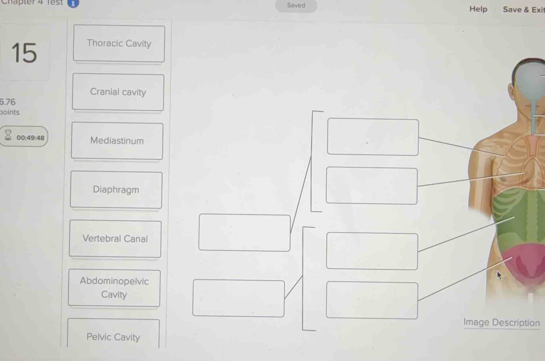
The diagram illustrates the subdivisions of the anterior/ventral body cavity. The two main boxes on the left represent the primary divisions of the ventral cavity: the upper division is the Thoracic Cavity and the lower division is the Abdominopelvic Cavity.
Subdivide the upper ventral cavity
The upper main box (Thoracic Cavity) branches into two subdivisions shown on the right:
- The upper subdivision pointing to the chest region is the Thoracic Cavity itself (or specifically the Mediastinum for the central portion containing the heart, while the overall upper cavity is the thoracic cavity). Looking at the labels, the top right box points to the chest area generally, representing the Thoracic Cavity, and the box below it points to the central heart region, representing the Mediastinum.
Subdivide the lower ventral cavity
The lower main box (Abdominopelvic Cavity) branches into two subdivisions:
- The upper subdivision pointing to the green abdominal region is the Abdominal Cavity (or the abdominal portion of the abdominopelvic cavity).
- The lower subdivision pointing to the pink pelvic region is the Pelvic Cavity.
Match labels to the diagram boxes
Let's align the draggable labels to the empty boxes from left to right, top to bottom:
- Left-middle upper box: Thoracic Cavity (which then branches into the mediastinum and pleural cavities).
- Left-middle lower box: Abdominopelvic Cavity (which branches into the abdominal and pelvic cavities).
- Top-right box (pointing to the chest/pleural area): Thoracic Cavity (or Mediastinum for the central chest). Let's look at the pointers:
- The top-right pointer goes to the upper chest area: Thoracic Cavity.
- The second-right pointer goes to the central mediastinum/heart area: Mediastinum.
- The third-right pointer goes to the green abdominal area: Abdominal Cavity (or Abdominopelvic Cavity as the parent, but here the subdivisions are abdominal and pelvic).
- The bottom-right pointer goes to the pink pelvic area: Pelvic Cavity.
Let's re-verify the standard anatomical hierarchy shown in this specific McGraw-Hill style diagram:
- Ventral Cavity is divided into:
- Thoracic Cavity (left upper box)
- Subdivisions: Mediastinum (top right box) and Pleural Cavity (not labeled here, or the top right box is Thoracic Cavity and the one below is Mediastinum). Actually, the bracket on the right groups the top two boxes under the left-upper box. The left-upper box is Thoracic Cavity. The bracketed subdivisions of the thoracic cavity are the Mediastinum and pleural/pericardial areas.
- Abdominopelvic Cavity (left lower box)
- Bracketed subdivisions: Abdominal Cavity (third right box) and Pelvic Cavity (bottom right box).

Snap & solve any problem in the app
Get step-by-step solutions on Sovi AI
Photo-based solutions with guided steps
Explore more problems and detailed explanations
Based on the anatomical hierarchy of the ventral body cavity, the correct labels for the empty boxes from left to right, top to bottom are:
- Left Upper Box: Thoracic Cavity
- Left Lower Box: Abdominopelvic Cavity
- Top Right Box (pointing to upper chest): Thoracic Cavity
- Second Right Box (pointing to central chest): Mediastinum
- Third Right Box (pointing to green abdomen): Abdominopelvic Cavity (or Abdominal Cavity)
- Bottom Right Box (pointing to pink pelvis): Pelvic Cavity