QUESTION IMAGE
Question
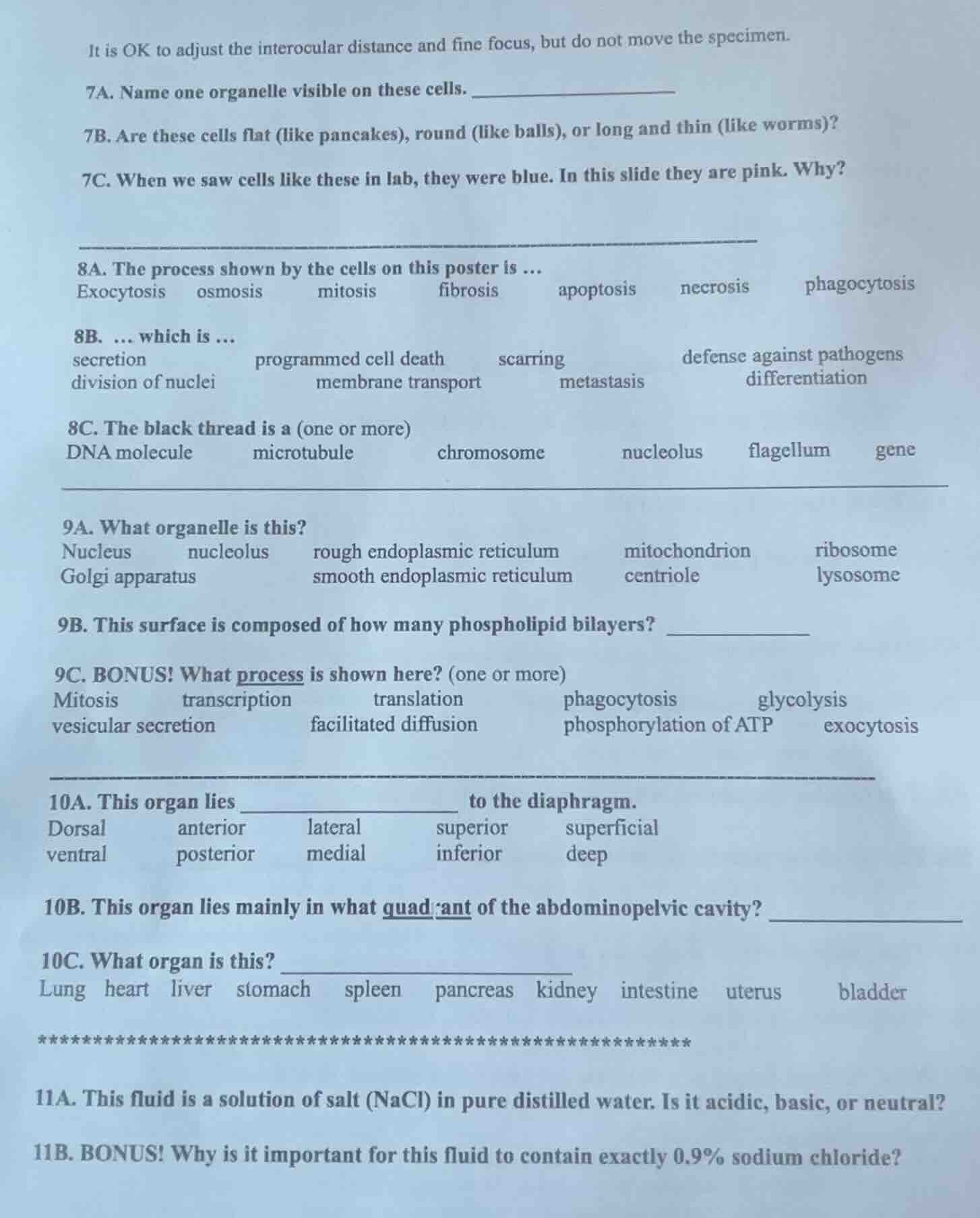
it is ok to adjust the interocular distance and fine focus, but do not move the specimen.
7a. name one organelle visible on these cells.
7b. are these cells flat (like pancakes), round (like balls), or long and thin (like worms)?
7c. when we saw cells like these in lab, they were blue. in this slide they are pink. why?
8a. the process shown by the cells on this poster is ...
exocytosis osmosis mitosis fibrosis apoptosis necrosis phagocytosis
8b. ... which is ...
secretion programmed cell death scarring defense against pathogens
division of nuclei membrane transport metastasis differentiation
8c. the black thread is a (one or more)
dna molecule microtubule chromosome nucleolus flagellum gene
9a. what organelle is this?
nucleus nucleolus rough endoplasmic reticulum mitochondrion ribosome
golgi apparatus smooth endoplasmic reticulum centriole lysosome
9b. this surface is composed of how many phospholipid bilayers?
9c. bonus! what process is shown here? (one or more)
mitosis transcription translation phagocytosis glycolysis
vesicular secretion facilitated diffusion phosphorylation of atp exocytosis
10a. this organ lies ______ to the diaphragm.
dorsal anterior lateral superior superficial
ventral posterior medial inferior deep
10b. this organ lies mainly in what quadrant of the abdominopelvic cavity?
10c. what organ is this?
lung heart liver stomach spleen pancreas kidney intestine uterus bladder
11a. this fluid is a solution of salt (nacl) in pure distilled water. is it acidic, basic, or neutral?
11b. bonus! why is it important for this fluid to contain exactly 0.9% sodium chloride?
Analyze questions 7A, 7B, and 7C (Microscope Specimen)
7A: The most prominent organelle visible in stained eukaryotic cells (like cheek cells or typical epithelial cells) under a light microscope is the nucleus.
7B: Epithelial cells (such as squamous cells) are flat (like pancakes).
7C: In lab, methylene blue is often used to stain cells blue. In this slide, they are pink because a different stain, such as eosin (part of the H&E stain), was used.
Analyze questions 8A, 8B, and 8C (Cell Division Poster)
8A: The cells on the poster show "mitosis" (cell division).
8B: Mitosis is the "division of nuclei".
8C: The "black thread" representing genetic material during mitosis is a "chromosome".
Analyze questions 9A, 9B, and 9C (Organelle Diagram)
9A: The organelle with a double membrane (composed of two phospholipid bilayers) and inner folds (cristae) is the "mitochondrion".
9B: The outer surface (envelope/membrane system) of a mitochondrion consists of an outer membrane and an inner membrane, which means it is composed of 2 phospholipid bilayers.
9C: The primary process shown/occurring here is the "phosphorylation of ATP" (cellular respiration).
Analyze questions 10A, 10B, and 10C (Organ Identification)
10C: Based on the options and standard anatomical models, the liver is a large organ situated immediately below the diaphragm.
10A: The liver lies "inferior" to the diaphragm.
10B: The liver lies mainly in the "right upper quadrant" (RUQ) of the abdominopelvic cavity.
Analyze questions 11A and 11B (Saline Solution)
11A: A solution of pure NaCl in distilled water is "neutral" (pH of approximately 7.0).
11B: A 0.9% NaCl solution is isotonic to human body fluids (red blood cells), preventing cells from shrinking (crenation) or swelling and bursting (lysis).

Snap & solve any problem in the app
Get step-by-step solutions on Sovi AI
Photo-based solutions with guided steps
Explore more problems and detailed explanations
Question 7A
Nucleus
Question 7B
flat (like pancakes)
Question 7C
A different stain (such as eosin) was used instead of methylene blue.
Question 8A
mitosis
Question 8B
division of nuclei
Question 8C
chromosome
Question 9A
mitochondrion
Question 9B
2
Question 9C
phosphorylation of ATP
Question 10A
inferior
Question 10B
right upper quadrant (RUQ)
Question 10C
liver
Question 11A
neutral
Question 11B
It is isotonic to body cells, preventing them from shrinking or swelling and bursting.