QUESTION IMAGE
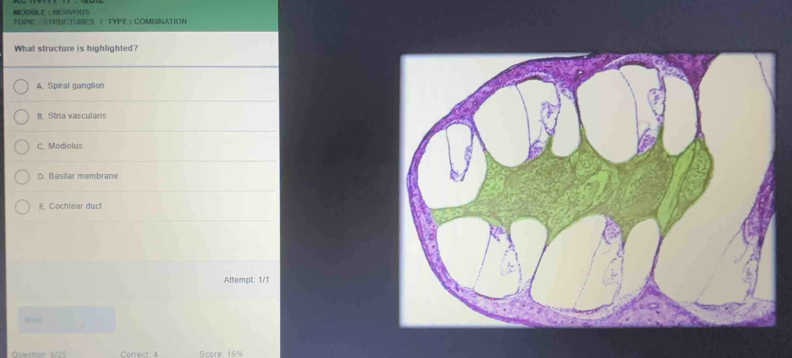
Question
module: nervous
topic: structures / type: combination
what structure is highlighted?
a. spiral ganglion
b. stria vascularis
c. modiolus
d. basilar membrane
e. cochlear duct
Identify the histological section
Using the Cochlea Histology Identification knowledge point
The provided image shows a cross-section of the bony cochlea of the inner ear, displaying its spiral turns around a central core.
Analyze the highlighted region
Using the Cochlea Histology Identification knowledge point
The green highlighted area is the central, conical pillar of spongy bone around which the cochlea wraps. This central bony core is known as the modiolus.
Evaluate the given options
Using the Cochlea Histology Identification knowledge point
- A. Spiral ganglion: Located within the modiolus, but the highlight covers the entire central bony core (modiolus) rather than just the neural cell bodies.
- B. Stria vascularis: Located on the outer wall of the cochlear duct.
- C. Modiolus: The central bony axis of the cochlea, which is highlighted in green.
- D. Basilar membrane: The floor of the cochlear duct supporting the organ of Corti.
- E. Cochlear duct: The middle fluid-filled chamber (scala media).

Snap & solve any problem in the app
Get step-by-step solutions on Sovi AI
Photo-based solutions with guided steps
Explore more problems and detailed explanations
- (A) Spiral ganglion
- (B) Stria vascularis
- (C) Modiolus (Correct answer)
- (D) Basilar membrane
- (E) Cochlear duct