QUESTION IMAGE
Question
module: nervous
topic: structures / type: combination
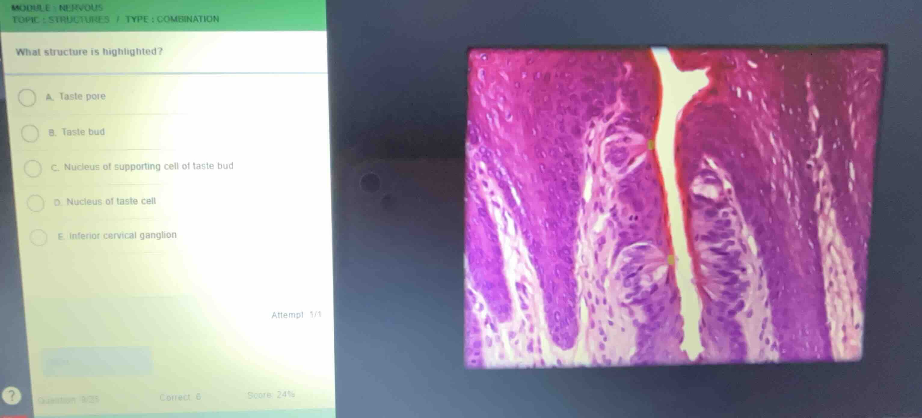
what structure is highlighted?
a. taste pore
b. taste bud
c. nucleus of supporting cell of taste bud
d. nucleus of taste cell
e. inferior cervical ganglion
attempt 1/1
Identify the tissue and structure
Using the Tongue Histology, Vallate Papilla, and Taste Buds knowledge points
- The histological section shows stratified squamous epithelium lining the trench of a vallate papilla on the tongue.
- Embedded within the lateral walls of the epithelium are pale, oval-shaped structures containing specialized sensory cells.
- These distinct, barrel-shaped structures are taste buds.
Determine the highlighted feature
- The question asks "What structure is highlighted?".
- Looking closely at the image, there is a small green highlight marker located at the apical opening of one of the taste buds, leading into the trench (the furrow).
- This small opening at the top of the taste bud through which microvilli project is the taste pore.

Snap & solve any problem in the app
Get step-by-step solutions on Sovi AI
Photo-based solutions with guided steps
Explore more problems and detailed explanations
- (A) Taste pore (Correct answer)
- (B) Taste bud
- (C) Nucleus of supporting cell of taste bud
- (D) Nucleus of taste cell
- (E) Inferior cervical ganglion