QUESTION IMAGE
Question
module: nervous
topic: structures / type: combination
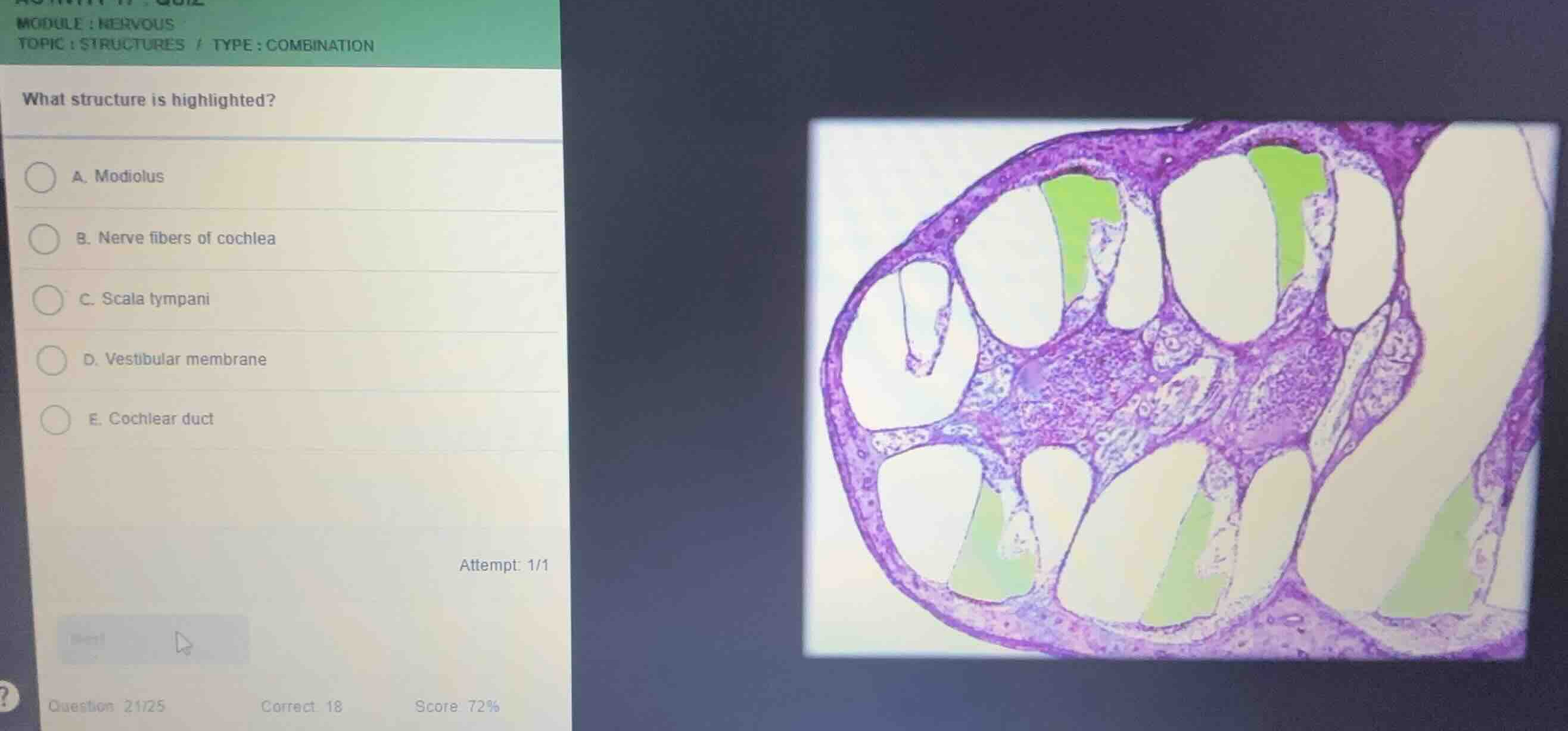
what structure is highlighted?
a. modiolus
b. nerve fibers of cochlea
c. scala tympani
d. vestibular membrane
e. cochlear duct
Identify the histological section
Using the Cochlea Histology knowledge point, we recognize this image as a cross-section of the cochlea, showing the spiral turns around the central bony hub (modiolus).
Analyze the three fluid-filled chambers
Using the Scala Vestibuli and Scala Tympani knowledge points, each turn of the cochlea contains three distinct ducts:
- Scala vestibuli: The upper chamber, filled with perilymph.
- Cochlear duct (Scala media): The middle triangular chamber, filled with endolymph.
- Scala tympani: The lower chamber, filled with perilymph.
Determine the highlighted structure
The green highlighted regions are located in the upper chamber of each cochlear turn, which is situated above the vestibular membrane and the cochlear duct. This upper chamber is the scala vestibuli (not listed as a direct option, but let's re-verify the orientation and options).
Wait, let's look closely at the orientation of the cochlear turns:
- The vestibular membrane separates the scala vestibuli from the cochlear duct.
- The basilar membrane separates the cochlear duct from the scala tympani.
- In this section, the vestibular membrane is the thin diagonal membrane on top of the cochlear duct, and the basilar membrane is the thicker membrane below it supporting the organ of Corti.
- The highlighted green spaces are located above the vestibular membrane, which corresponds to the scala vestibuli. However, looking at the options:
- A. Modiolus
- B. Nerve fibers of cochlea
- C. Scala tympani
- D. Vestibular membrane
- E. Cochlear duct
- Let's re-examine the orientation. If the highlighted chambers are at the bottom in some turns and top in others, let's trace the organ of Corti.
- The organ of Corti sits on the basilar membrane. The basilar membrane is the floor of the cochlear duct, separating it from the scala tympani.
- In the turns shown:
- The middle chamber containing the organ of Corti is the cochlear duct.
- The chamber below the basilar membrane (where the organ of Corti sits) is the scala tympani.
- Let's check the position of the green highlights relative to the organ of Corti:
- In the upper turns, the green highlight is on the side opposite the basilar membrane, which is the scala vestibuli.
- Wait, let's look at the lower turns: the green highlights are at the bottom.
- Let's trace the vestibular membrane (thin) and basilar membrane (thicker, with organ of Corti).
- Actually, the chamber highlighted in green is always on the side of the vestibular membrane (upper chamber, scala vestibuli) or is it the scala tympani?
- Let's look at the options again. Since "Scala vestibuli" is NOT an option, let's double check if the orientation is inverted, making the highlighted region the scala tympani (Option C).
- In many standard histology slides of the cochlea, the scala vestibuli is on top and the scala tympani is at the bottom. If the slide is oriented such that the scala tympani is highlighted, let's verify: the scala tympani is the lower compartment. In this image, the green highl…

Snap & solve any problem in the app
Get step-by-step solutions on Sovi AI
Photo-based solutions with guided steps
Explore more problems and detailed explanations
- (A) Modiolus
- (B) Nerve fibers of cochlea
- (C) Scala tympani (Correct answer)
- (D) Vestibular membrane
- (E) Cochlear duct