QUESTION IMAGE
Question
mitochondrion
vesicle
cytoskeleton
rough er
cytoplasm
nuclear envelope
centrioles (in centrosome)
nucleolus
chromatin
lysosome
nuclear pore
Identify the mislabeled organelles
Using the Mitochondria Function knowledge point
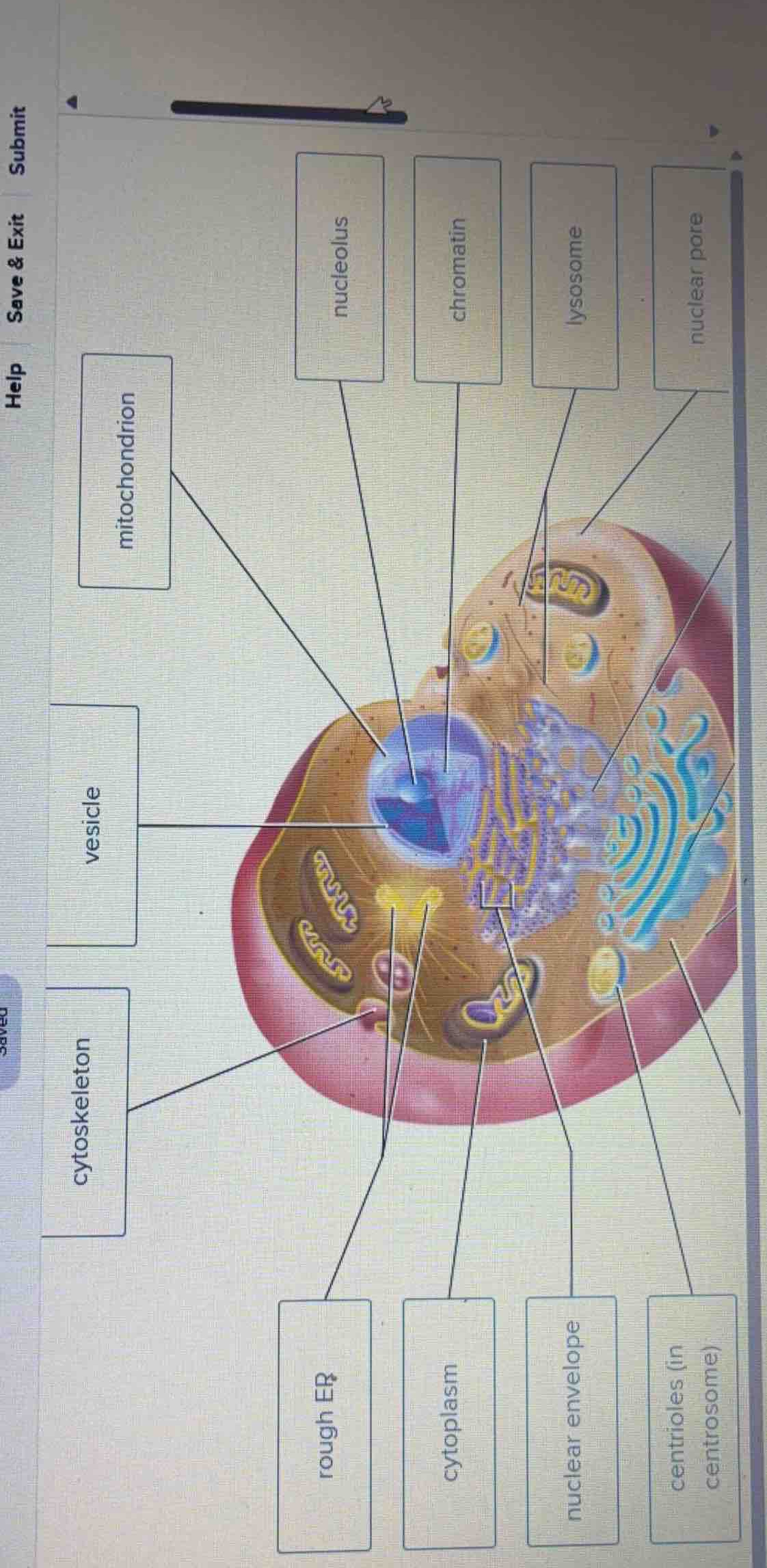
The diagram shows a 3D cutaway of an animal cell with several labels pointing to various organelles and cellular structures. Let's analyze the pointer lines for the labels that are currently filled in to see if they are pointing to the correct structures:
- mitochondrion: The pointer line for this box points directly to the nucleus (specifically the outer nuclear region/nucleoplasm), not a mitochondrion. This label is incorrect.
- vesicle: The pointer line for this box points to a small spherical structure near the nucleus, which is indeed a vesicle. This label is correct.
- cytoskeleton: The pointer line for this box points to the microfilaments/microtubules (fibers) in the cytoplasm, which represents the cytoskeleton. This label is correct.
- rough ER: The pointer line for this box points to the yellow-brown tubular network with yellow dots (ribosomes) on it, which is the rough endoplasmic reticulum (rough ER). This label is correct.
- cytoplasm: The pointer line for this box points to the fluid-filled space of the cell, which is the cytoplasm. This label is correct.
- nuclear envelope: The pointer line for this box points to the double membrane surrounding the nucleus, which is the nuclear envelope. This label is correct.
- centrioles (in centrosome): The pointer line for this box points to the yellow cylindrical structures near the nucleus, which are the centrioles. This label is correct.
Determine correct placements for empty labels
Now let's look at the empty boxes on the right and top-right, and find where their corresponding structures are located:
- nucleolus: This should point to the dark, dense spherical region inside the nucleus.
- chromatin: This should point to the thread-like DNA/protein fibers inside the nucleoplasm.
- lysosome: This should point to a membrane-bound organelle containing digestive enzymes (often depicted as a spherical vesicle with internal material).
- nuclear pore: This should point to the small openings/channels in the nuclear envelope.
Looking closely at the image, the box labeled mitochondrion is pointing to the nucleus, which is incorrect. The actual mitochondria are the oval-shaped organelles with folded inner membranes (cristae) shown in orange/brown scattered in the cytoplasm.
To complete the labeling activity correctly:
- The label mitochondrion needs to be moved to point to one of the orange, oval-shaped organelles with folded inner membranes (cristae) located in the cytoplasm.
- The box currently labeled mitochondrion (pointing to the nucleus) should be replaced with the correct nuclear component label or the label for the nucleus itself.

Snap & solve any problem in the app
Get step-by-step solutions on Sovi AI
Photo-based solutions with guided steps
Explore more problems and detailed explanations
Based on the visual analysis of the cell diagram, the labels should be placed as follows:
- Mislabeled Item:
- The box labeled mitochondrion is currently pointing incorrectly to the nucleus. It should be moved to point to one of the orange, oval-shaped organelles with folded inner membranes (cristae) located in the cytoplasm.
- Correct Placements for the Remaining Labels:
- nucleolus: Points to the dark, dense spherical region at the very center of the nucleus.
- chromatin: Points to the granular, thread-like material inside the nucleus surrounding the nucleolus.
- nuclear pore: Points to the small circular openings on the surface of the nuclear envelope.
- lysosome: Points to the spherical membrane-bound organelles in the cytoplasm containing digestive enzymes (distinct from simple transport vesicles).