QUESTION IMAGE
Question
Question was provided via image upload.
Identify the anatomical structure
Using the Eye Anatomy and Ocular Layers knowledge points
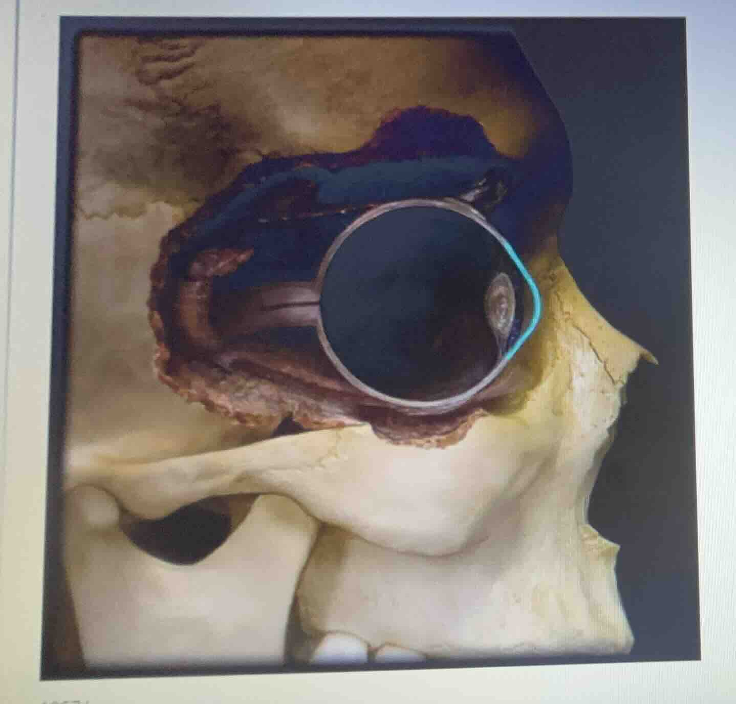
The image shows a sagittal/cross-sectional anatomical model of the human eye situated within the bony orbit of the skull.
Analyze the highlighted component
Using the Optical Components and Lens Structure knowledge points
The light blue/cyan highlight outlines the anterior, transparent, curved outer layer at the front of the eyeball, which is the cornea.
Identify the posterior structures
Using the Neural Components and Retina Anatomy knowledge points
The model also depicts the internal chamber, the lens situated behind the pupil, and the optic nerve exiting the posterior aspect of the globe.

Snap & solve any problem in the app
Get step-by-step solutions on Sovi AI
Photo-based solutions with guided steps
Explore more problems and detailed explanations
This anatomical model depicts a cross-section of the human eye (eyeball) situated within the bony orbit (orbital cavity) of the skull.
Key structures visible in this model include:
- Cornea: Highlighted by the light blue outline at the anterior surface.
- Lens: The transparent biconvex structure located just behind the pupil/iris.
- Optic Nerve: The cord-like structure extending from the posterior aspect of the eye toward the brain.
- Sclera/Retina Layers: The outer protective coat and inner neural layers forming the wall of the globe.