QUESTION IMAGE
Question
activity 17 : quiz
module : nervous
topic : structures / type : combination
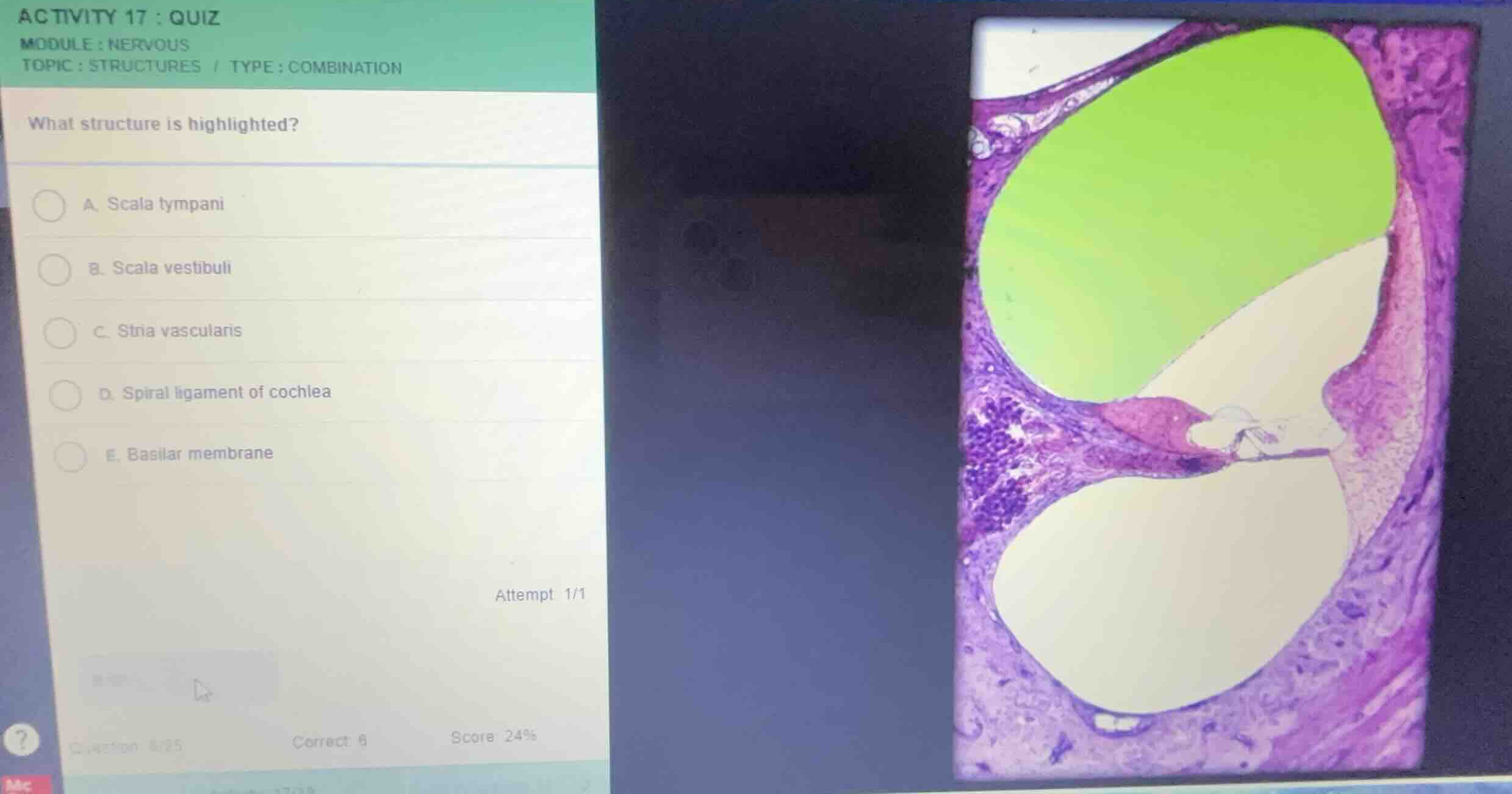
what structure is highlighted?
a. scala tympani
b. scala vestibuli
c. stria vascularis
d. spiral ligament of cochlea
e. basilar membrane
Identify the anatomical orientation of the cochlea
Using the Cochlea Histology knowledge point
The image shows a cross-section of a turn of the cochlea. The cochlea is divided into three fluid-filled chambers: the upper chamber, the middle chamber (cochlear duct or scala media), and the lower chamber.
Locate the highlighted chamber
Using the Scala Vestibuli knowledge point
The highlighted region in green is the uppermost chamber of the cochlear duct system. This chamber is located superior to the vestibular membrane (Reissner's membrane) and is filled with perilymph. This upper chamber is anatomically identified as the scala vestibuli.

Snap & solve any problem in the app
Get step-by-step solutions on Sovi AI
Photo-based solutions with guided steps
Explore more problems and detailed explanations
- A. Scala tympani
- B. Scala vestibuli (Correct answer)
- C. Stria vascularis
- D. Spiral ligament of cochlea
- E. Basilar membrane